Zinc »
PDB 5llp-5lsy »
5lrm »
Zinc in PDB 5lrm: Structure of Di-Zinc Mcr-1 in P41212 Space Group
Protein crystallography data
The structure of Structure of Di-Zinc Mcr-1 in P41212 Space Group, PDB code: 5lrm
was solved by
P.Hinchliffe,
J.Spencer,
with X-Ray Crystallography technique. A brief refinement statistics is given in the table below:
| Resolution Low / High (Å) | 48.09 / 1.75 |
| Space group | P 41 21 2 |
| Cell size a, b, c (Å), α, β, γ (°) | 49.044, 49.044, 244.253, 90.00, 90.00, 90.00 |
| R / Rfree (%) | 16.9 / 19.8 |
Zinc Binding Sites:
The binding sites of Zinc atom in the Structure of Di-Zinc Mcr-1 in P41212 Space Group
(pdb code 5lrm). This binding sites where shown within
5.0 Angstroms radius around Zinc atom.
In total 2 binding sites of Zinc where determined in the Structure of Di-Zinc Mcr-1 in P41212 Space Group, PDB code: 5lrm:
Jump to Zinc binding site number: 1; 2;
In total 2 binding sites of Zinc where determined in the Structure of Di-Zinc Mcr-1 in P41212 Space Group, PDB code: 5lrm:
Jump to Zinc binding site number: 1; 2;
Zinc binding site 1 out of 2 in 5lrm
Go back to
Zinc binding site 1 out
of 2 in the Structure of Di-Zinc Mcr-1 in P41212 Space Group
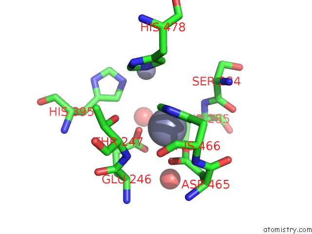
Mono view
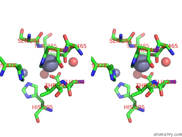
Stereo pair view
Mono view
Stereo pair view
A full contact list of Zinc with other atoms in the Zn binding
site number 1 of Structure of Di-Zinc Mcr-1 in P41212 Space Group within 5.0Å range:
|
Zinc binding site 2 out of 2 in 5lrm
Go back to
Zinc binding site 2 out
of 2 in the Structure of Di-Zinc Mcr-1 in P41212 Space Group
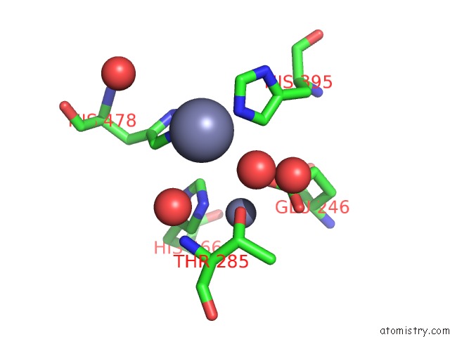
Mono view
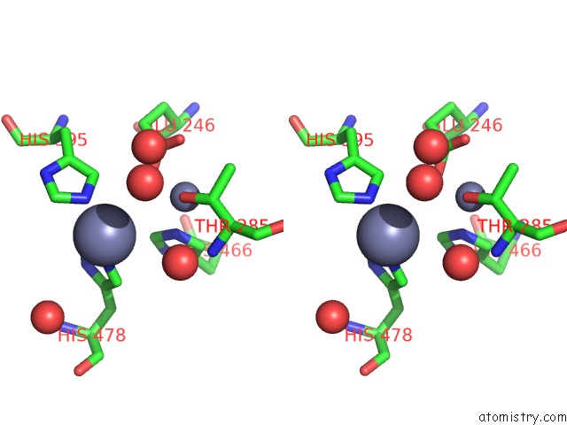
Stereo pair view
Mono view
Stereo pair view
A full contact list of Zinc with other atoms in the Zn binding
site number 2 of Structure of Di-Zinc Mcr-1 in P41212 Space Group within 5.0Å range:
|
Reference:
P.Hinchliffe,
Q.E.Yang,
E.Portal,
T.Young,
H.Li,
C.L.Tooke,
M.J.Carvalho,
N.G.Paterson,
J.Brem,
P.R.Niumsup,
U.Tansawai,
L.Lei,
M.Li,
Z.Shen,
Y.Wang,
C.J.Schofield,
A.J.Mulholland,
J.Shen,
N.Fey,
T.R.Walsh,
J.Spencer.
Insights Into the Mechanistic Basis of Plasmid-Mediated Colistin Resistance From Crystal Structures of the Catalytic Domain of Mcr-1. Sci Rep V. 7 39392 2017.
ISSN: ESSN 2045-2322
PubMed: 28059088
DOI: 10.1038/SREP39392
Page generated: Sun Oct 27 21:16:26 2024
ISSN: ESSN 2045-2322
PubMed: 28059088
DOI: 10.1038/SREP39392
Last articles
Zn in 9MJ5Zn in 9HNW
Zn in 9G0L
Zn in 9FNE
Zn in 9DZN
Zn in 9E0I
Zn in 9D32
Zn in 9DAK
Zn in 8ZXC
Zn in 8ZUF