Zinc »
PDB 5lcf-5llo »
5lhd »
Zinc in PDB 5lhd: Structure of Glycosylated Human Aminopeptidase N
Enzymatic activity of Structure of Glycosylated Human Aminopeptidase N
All present enzymatic activity of Structure of Glycosylated Human Aminopeptidase N:
3.4.11.2;
3.4.11.2;
Protein crystallography data
The structure of Structure of Glycosylated Human Aminopeptidase N, PDB code: 5lhd
was solved by
R.Recacha,
G.Mudgal,
C.Santiago,
J.M.Casasnovas,
with X-Ray Crystallography technique. A brief refinement statistics is given in the table below:
| Resolution Low / High (Å) | 19.97 / 2.60 |
| Space group | P 21 21 21 |
| Cell size a, b, c (Å), α, β, γ (°) | 127.093, 168.856, 244.249, 90.00, 90.00, 90.00 |
| R / Rfree (%) | 18.2 / 21.4 |
Zinc Binding Sites:
The binding sites of Zinc atom in the Structure of Glycosylated Human Aminopeptidase N
(pdb code 5lhd). This binding sites where shown within
5.0 Angstroms radius around Zinc atom.
In total 4 binding sites of Zinc where determined in the Structure of Glycosylated Human Aminopeptidase N, PDB code: 5lhd:
Jump to Zinc binding site number: 1; 2; 3; 4;
In total 4 binding sites of Zinc where determined in the Structure of Glycosylated Human Aminopeptidase N, PDB code: 5lhd:
Jump to Zinc binding site number: 1; 2; 3; 4;
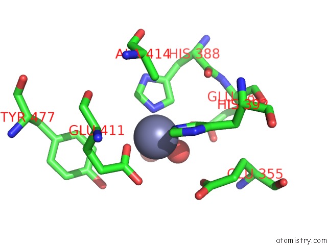
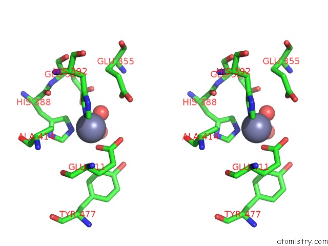
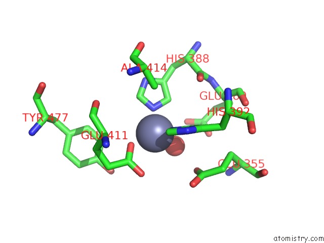
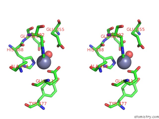
Zinc binding site 1 out of 4 in 5lhd
Go back to
Zinc binding site 1 out
of 4 in the Structure of Glycosylated Human Aminopeptidase N
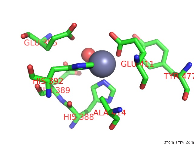
Mono view
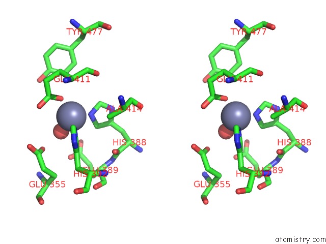
Stereo pair view
Mono view
Stereo pair view
A full contact list of Zinc with other atoms in the Zn binding
site number 1 of Structure of Glycosylated Human Aminopeptidase N within 5.0Å range:
|
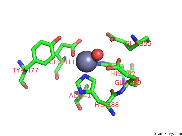
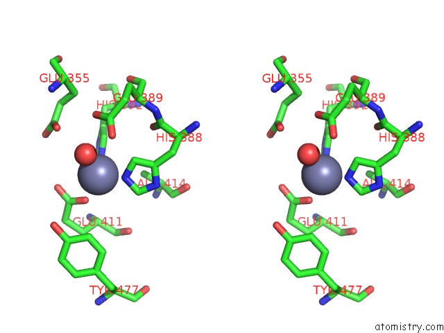
Zinc binding site 2 out of 4 in 5lhd
Go back to
Zinc binding site 2 out
of 4 in the Structure of Glycosylated Human Aminopeptidase N
Mono view
Stereo pair view
Mono view
Stereo pair view
A full contact list of Zinc with other atoms in the Zn binding
site number 2 of Structure of Glycosylated Human Aminopeptidase N within 5.0Å range:
|
Zinc binding site 3 out of 4 in 5lhd
Go back to
Zinc binding site 3 out
of 4 in the Structure of Glycosylated Human Aminopeptidase N
Mono view
Stereo pair view
Mono view
Stereo pair view
A full contact list of Zinc with other atoms in the Zn binding
site number 3 of Structure of Glycosylated Human Aminopeptidase N within 5.0Å range:
|
Zinc binding site 4 out of 4 in 5lhd
Go back to
Zinc binding site 4 out
of 4 in the Structure of Glycosylated Human Aminopeptidase N
Mono view
Stereo pair view
Mono view
Stereo pair view
A full contact list of Zinc with other atoms in the Zn binding
site number 4 of Structure of Glycosylated Human Aminopeptidase N within 5.0Å range:
|
Reference:
C.Santiago,
G.Mudgal,
J.Reguera,
R.Recacha,
S.Albrecht,
L.Enjuanes,
J.M.Casasnovas.
Allosteric Inhibition of Aminopeptidase N Functions Related to Tumor Growth and Virus Infection. Sci Rep V. 7 46045 2017.
ISSN: ESSN 2045-2322
PubMed: 28393915
DOI: 10.1038/SREP46045
Page generated: Sun Oct 27 21:01:28 2024
ISSN: ESSN 2045-2322
PubMed: 28393915
DOI: 10.1038/SREP46045
Last articles
Zn in 9MJ5Zn in 9HNW
Zn in 9G0L
Zn in 9FNE
Zn in 9DZN
Zn in 9E0I
Zn in 9D32
Zn in 9DAK
Zn in 8ZXC
Zn in 8ZUF