Zinc »
PDB 5l0e-5lca »
5lbz »
Zinc in PDB 5lbz: Structure of the Human Quinone Reductase 2 (NQO2) in Complex with Pacritinib
Enzymatic activity of Structure of the Human Quinone Reductase 2 (NQO2) in Complex with Pacritinib
All present enzymatic activity of Structure of the Human Quinone Reductase 2 (NQO2) in Complex with Pacritinib:
1.10.5.1;
1.10.5.1;
Protein crystallography data
The structure of Structure of the Human Quinone Reductase 2 (NQO2) in Complex with Pacritinib, PDB code: 5lbz
was solved by
S.Schneider,
G.Medard,
B.Kuster,
with X-Ray Crystallography technique. A brief refinement statistics is given in the table below:
| Resolution Low / High (Å) | 48.60 / 1.40 |
| Space group | P 21 21 21 |
| Cell size a, b, c (Å), α, β, γ (°) | 61.454, 79.220, 106.230, 90.00, 90.00, 90.00 |
| R / Rfree (%) | 10.9 / 13.1 |
Zinc Binding Sites:
The binding sites of Zinc atom in the Structure of the Human Quinone Reductase 2 (NQO2) in Complex with Pacritinib
(pdb code 5lbz). This binding sites where shown within
5.0 Angstroms radius around Zinc atom.
In total 2 binding sites of Zinc where determined in the Structure of the Human Quinone Reductase 2 (NQO2) in Complex with Pacritinib, PDB code: 5lbz:
Jump to Zinc binding site number: 1; 2;
In total 2 binding sites of Zinc where determined in the Structure of the Human Quinone Reductase 2 (NQO2) in Complex with Pacritinib, PDB code: 5lbz:
Jump to Zinc binding site number: 1; 2;
Zinc binding site 1 out of 2 in 5lbz
Go back to
Zinc binding site 1 out
of 2 in the Structure of the Human Quinone Reductase 2 (NQO2) in Complex with Pacritinib
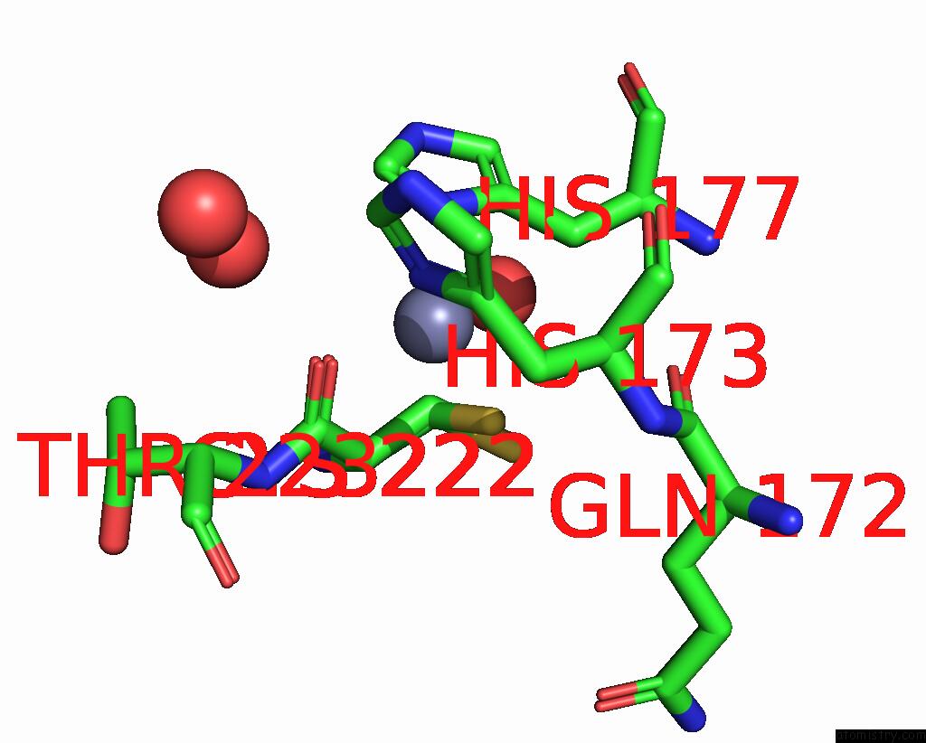
Mono view
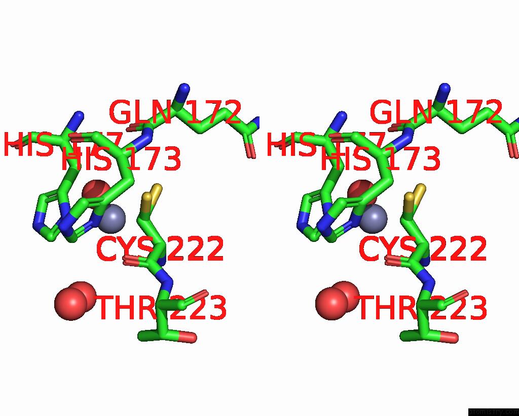
Stereo pair view
Mono view
Stereo pair view
A full contact list of Zinc with other atoms in the Zn binding
site number 1 of Structure of the Human Quinone Reductase 2 (NQO2) in Complex with Pacritinib within 5.0Å range:
|
Zinc binding site 2 out of 2 in 5lbz
Go back to
Zinc binding site 2 out
of 2 in the Structure of the Human Quinone Reductase 2 (NQO2) in Complex with Pacritinib
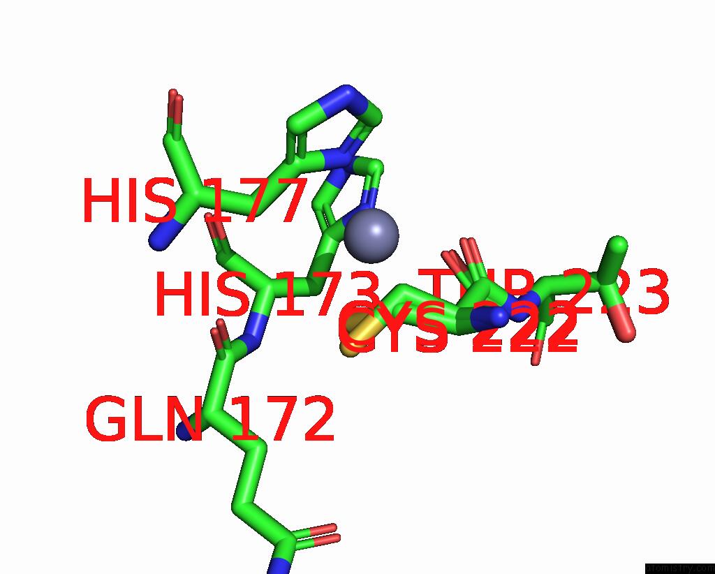
Mono view
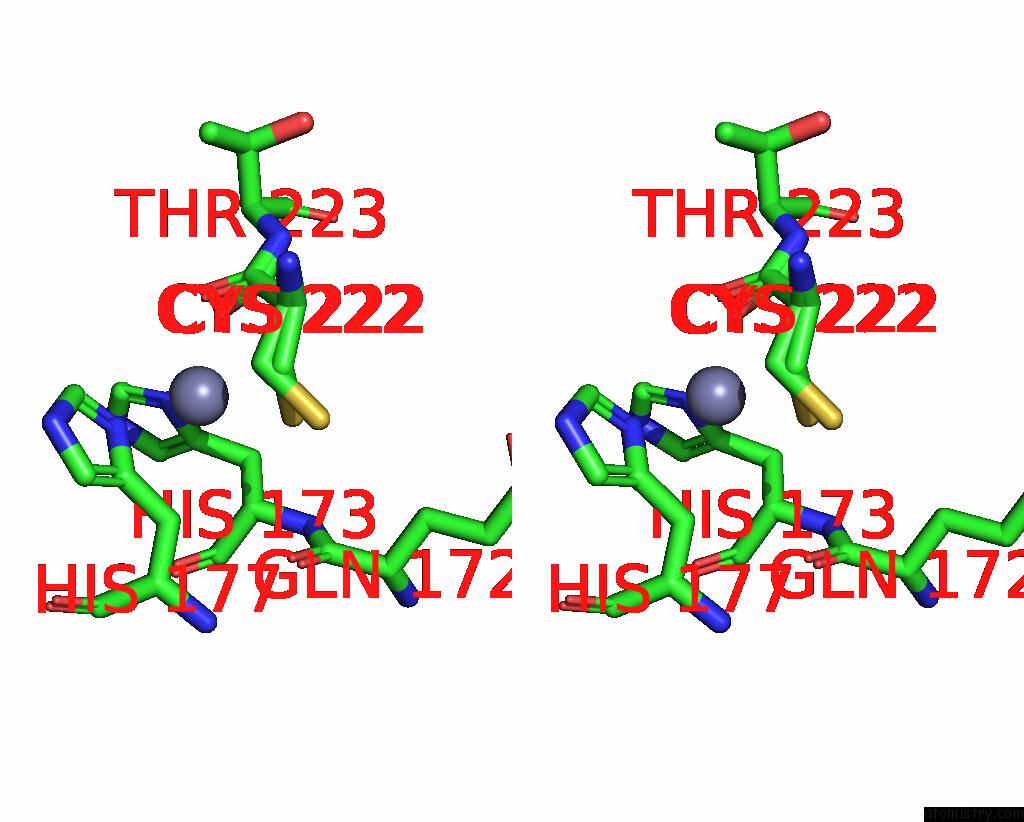
Stereo pair view
Mono view
Stereo pair view
A full contact list of Zinc with other atoms in the Zn binding
site number 2 of Structure of the Human Quinone Reductase 2 (NQO2) in Complex with Pacritinib within 5.0Å range:
|
Reference:
S.Klaeger,
S.Heinzlmeir,
M.Wilhelm,
H.Polzer,
B.Vick,
P.A.Koenig,
M.Reinecke,
B.Ruprecht,
S.Petzoldt,
C.Meng,
J.Zecha,
K.Reiter,
H.Qiao,
D.Helm,
H.Koch,
M.Schoof,
G.Canevari,
E.Casale,
S.R.Depaolini,
A.Feuchtinger,
Z.Wu,
T.Schmidt,
L.Rueckert,
W.Becker,
J.Huenges,
A.K.Garz,
B.O.Gohlke,
D.P.Zolg,
G.Kayser,
T.Vooder,
R.Preissner,
H.Hahne,
N.Tonisson,
K.Kramer,
K.Gotze,
F.Bassermann,
J.Schlegl,
H.C.Ehrlich,
S.Aiche,
A.Walch,
P.A.Greif,
S.Schneider,
E.R.Felder,
J.Ruland,
G.Medard,
I.Jeremias,
K.Spiekermann,
B.Kuster.
The Target Landscape of Clinical Kinase Drugs. Science V. 358 2017.
ISSN: ESSN 1095-9203
PubMed: 29191878
DOI: 10.1126/SCIENCE.AAN4368
Page generated: Sun Oct 27 20:55:46 2024
ISSN: ESSN 1095-9203
PubMed: 29191878
DOI: 10.1126/SCIENCE.AAN4368
Last articles
Zn in 9MJ5Zn in 9HNW
Zn in 9G0L
Zn in 9FNE
Zn in 9DZN
Zn in 9E0I
Zn in 9D32
Zn in 9DAK
Zn in 8ZXC
Zn in 8ZUF