Zinc »
PDB 5j1q-5jf1 »
5jex »
Zinc in PDB 5jex: Crystal Structure of Type 2 Pdf From Streptococcus Agalactiae, Crystallized in Imidazole Buffer
Enzymatic activity of Crystal Structure of Type 2 Pdf From Streptococcus Agalactiae, Crystallized in Imidazole Buffer
All present enzymatic activity of Crystal Structure of Type 2 Pdf From Streptococcus Agalactiae, Crystallized in Imidazole Buffer:
3.5.1.88;
3.5.1.88;
Protein crystallography data
The structure of Crystal Structure of Type 2 Pdf From Streptococcus Agalactiae, Crystallized in Imidazole Buffer, PDB code: 5jex
was solved by
S.Fieulaine,
C.Giglione,
T.Meinnel,
with X-Ray Crystallography technique. A brief refinement statistics is given in the table below:
| Resolution Low / High (Å) | 44.37 / 2.00 |
| Space group | P 21 21 21 |
| Cell size a, b, c (Å), α, β, γ (°) | 41.320, 65.550, 88.710, 90.00, 90.00, 90.00 |
| R / Rfree (%) | 15.9 / 20.8 |
Zinc Binding Sites:
The binding sites of Zinc atom in the Crystal Structure of Type 2 Pdf From Streptococcus Agalactiae, Crystallized in Imidazole Buffer
(pdb code 5jex). This binding sites where shown within
5.0 Angstroms radius around Zinc atom.
In total 9 binding sites of Zinc where determined in the Crystal Structure of Type 2 Pdf From Streptococcus Agalactiae, Crystallized in Imidazole Buffer, PDB code: 5jex:
Jump to Zinc binding site number: 1; 2; 3; 4; 5; 6; 7; 8; 9;
In total 9 binding sites of Zinc where determined in the Crystal Structure of Type 2 Pdf From Streptococcus Agalactiae, Crystallized in Imidazole Buffer, PDB code: 5jex:
Jump to Zinc binding site number: 1; 2; 3; 4; 5; 6; 7; 8; 9;
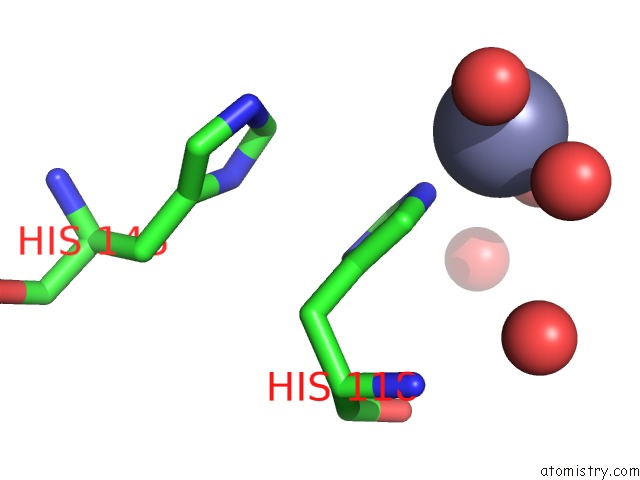
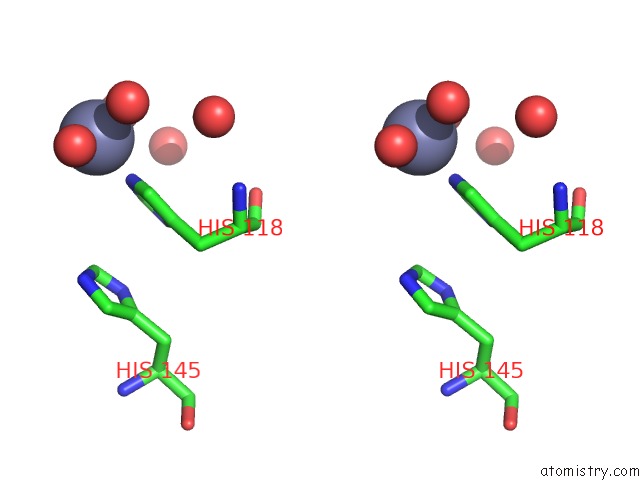
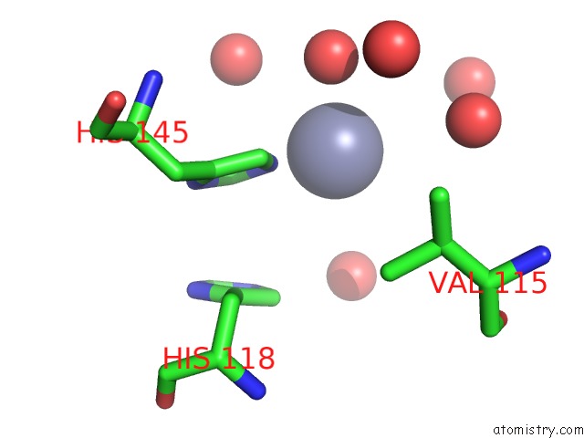
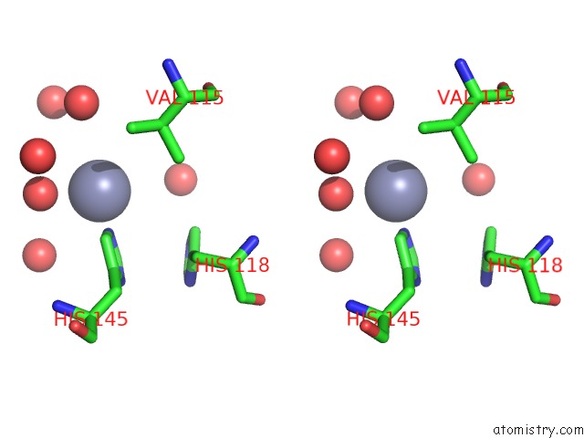
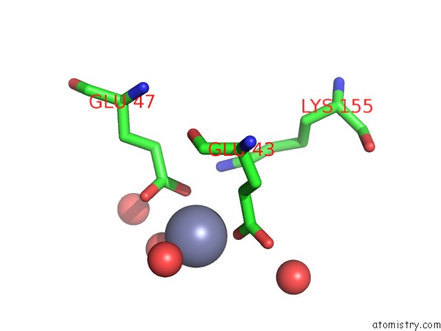
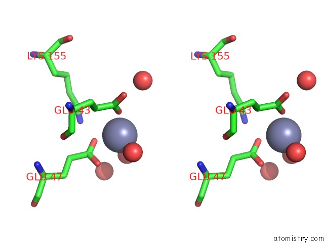
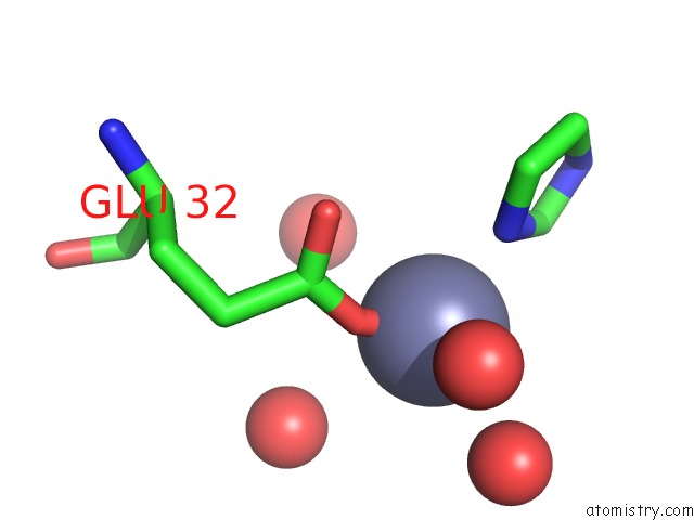
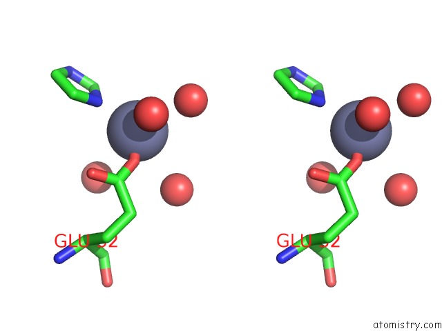
Zinc binding site 1 out of 9 in 5jex
Go back to
Zinc binding site 1 out
of 9 in the Crystal Structure of Type 2 Pdf From Streptococcus Agalactiae, Crystallized in Imidazole Buffer
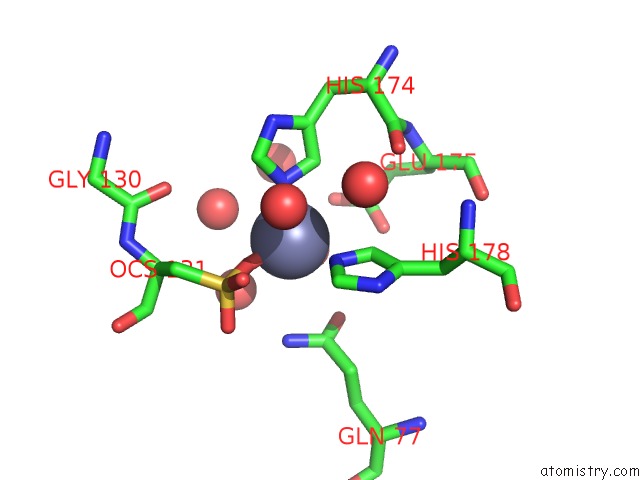
Mono view
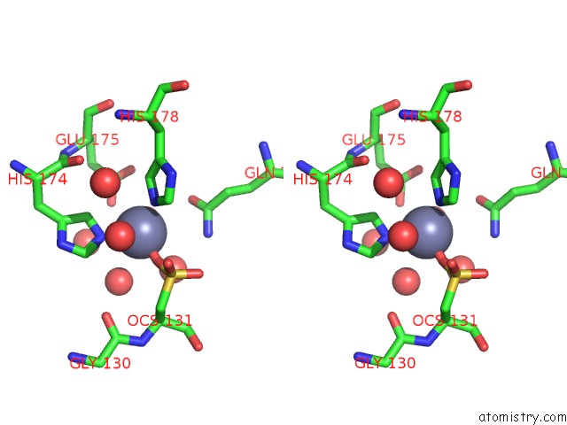
Stereo pair view
Mono view
Stereo pair view
A full contact list of Zinc with other atoms in the Zn binding
site number 1 of Crystal Structure of Type 2 Pdf From Streptococcus Agalactiae, Crystallized in Imidazole Buffer within 5.0Å range:
|
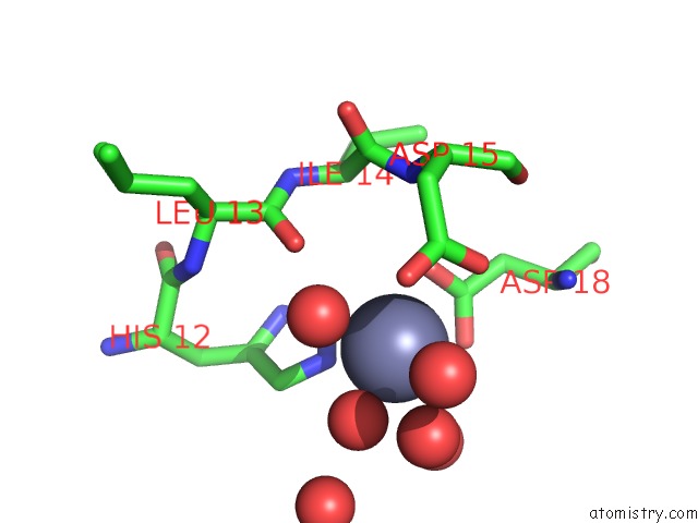
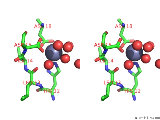
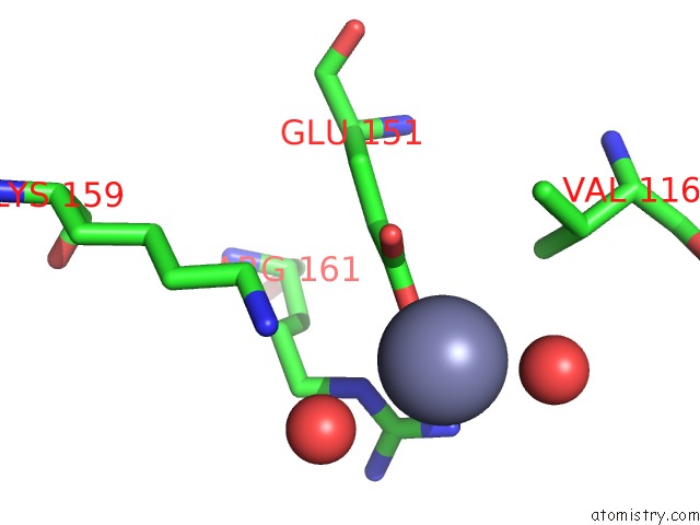
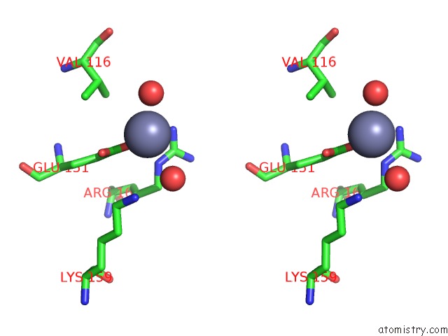
Zinc binding site 2 out of 9 in 5jex
Go back to
Zinc binding site 2 out
of 9 in the Crystal Structure of Type 2 Pdf From Streptococcus Agalactiae, Crystallized in Imidazole Buffer
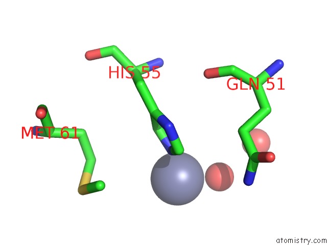
Mono view
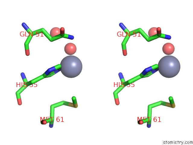
Stereo pair view
Mono view
Stereo pair view
A full contact list of Zinc with other atoms in the Zn binding
site number 2 of Crystal Structure of Type 2 Pdf From Streptococcus Agalactiae, Crystallized in Imidazole Buffer within 5.0Å range:
|
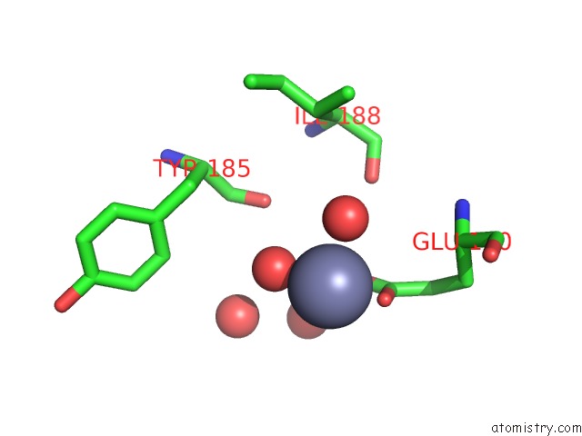
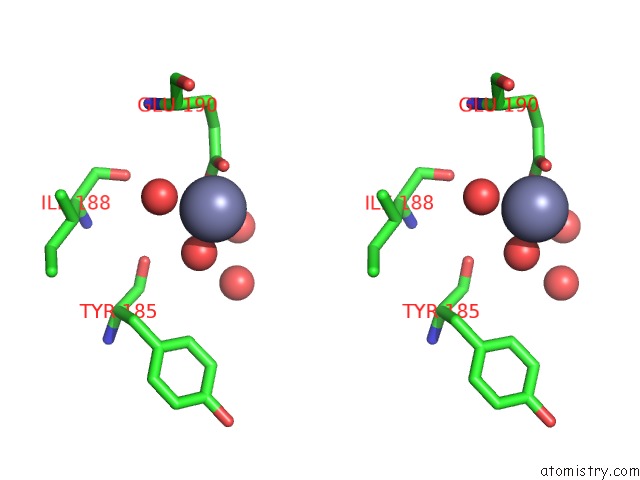
Zinc binding site 3 out of 9 in 5jex
Go back to
Zinc binding site 3 out
of 9 in the Crystal Structure of Type 2 Pdf From Streptococcus Agalactiae, Crystallized in Imidazole Buffer
Mono view
Stereo pair view
Mono view
Stereo pair view
A full contact list of Zinc with other atoms in the Zn binding
site number 3 of Crystal Structure of Type 2 Pdf From Streptococcus Agalactiae, Crystallized in Imidazole Buffer within 5.0Å range:
|
Zinc binding site 4 out of 9 in 5jex
Go back to
Zinc binding site 4 out
of 9 in the Crystal Structure of Type 2 Pdf From Streptococcus Agalactiae, Crystallized in Imidazole Buffer
Mono view
Stereo pair view
Mono view
Stereo pair view
A full contact list of Zinc with other atoms in the Zn binding
site number 4 of Crystal Structure of Type 2 Pdf From Streptococcus Agalactiae, Crystallized in Imidazole Buffer within 5.0Å range:
|
Zinc binding site 5 out of 9 in 5jex
Go back to
Zinc binding site 5 out
of 9 in the Crystal Structure of Type 2 Pdf From Streptococcus Agalactiae, Crystallized in Imidazole Buffer
Mono view
Stereo pair view
Mono view
Stereo pair view
A full contact list of Zinc with other atoms in the Zn binding
site number 5 of Crystal Structure of Type 2 Pdf From Streptococcus Agalactiae, Crystallized in Imidazole Buffer within 5.0Å range:
|
Zinc binding site 6 out of 9 in 5jex
Go back to
Zinc binding site 6 out
of 9 in the Crystal Structure of Type 2 Pdf From Streptococcus Agalactiae, Crystallized in Imidazole Buffer
Mono view
Stereo pair view
Mono view
Stereo pair view
A full contact list of Zinc with other atoms in the Zn binding
site number 6 of Crystal Structure of Type 2 Pdf From Streptococcus Agalactiae, Crystallized in Imidazole Buffer within 5.0Å range:
|
Zinc binding site 7 out of 9 in 5jex
Go back to
Zinc binding site 7 out
of 9 in the Crystal Structure of Type 2 Pdf From Streptococcus Agalactiae, Crystallized in Imidazole Buffer
Mono view
Stereo pair view
Mono view
Stereo pair view
A full contact list of Zinc with other atoms in the Zn binding
site number 7 of Crystal Structure of Type 2 Pdf From Streptococcus Agalactiae, Crystallized in Imidazole Buffer within 5.0Å range:
|
Zinc binding site 8 out of 9 in 5jex
Go back to
Zinc binding site 8 out
of 9 in the Crystal Structure of Type 2 Pdf From Streptococcus Agalactiae, Crystallized in Imidazole Buffer
Mono view
Stereo pair view
Mono view
Stereo pair view
A full contact list of Zinc with other atoms in the Zn binding
site number 8 of Crystal Structure of Type 2 Pdf From Streptococcus Agalactiae, Crystallized in Imidazole Buffer within 5.0Å range:
|
Zinc binding site 9 out of 9 in 5jex
Go back to
Zinc binding site 9 out
of 9 in the Crystal Structure of Type 2 Pdf From Streptococcus Agalactiae, Crystallized in Imidazole Buffer
Mono view
Stereo pair view
Mono view
Stereo pair view
A full contact list of Zinc with other atoms in the Zn binding
site number 9 of Crystal Structure of Type 2 Pdf From Streptococcus Agalactiae, Crystallized in Imidazole Buffer within 5.0Å range:
|
Reference:
S.Fieulaine,
R.Alves De Sousa,
L.Maigre,
K.Hamiche,
M.Alimi,
J.M.Bolla,
A.Taleb,
A.Denis,
J.M.Pages,
I.Artaud,
T.Meinnel,
C.Giglione.
A Unique Peptide Deformylase Platform to Rationally Design and Challenge Novel Active Compounds. Sci Rep V. 6 35429 2016.
ISSN: ESSN 2045-2322
PubMed: 27762275
DOI: 10.1038/SREP35429
Page generated: Sun Oct 27 18:46:54 2024
ISSN: ESSN 2045-2322
PubMed: 27762275
DOI: 10.1038/SREP35429
Last articles
Zn in 9MJ5Zn in 9HNW
Zn in 9G0L
Zn in 9FNE
Zn in 9DZN
Zn in 9E0I
Zn in 9D32
Zn in 9DAK
Zn in 8ZXC
Zn in 8ZUF