Zinc »
PDB 5ikf-5j1m »
5iye »
Zinc in PDB 5iye: Comparison of X-Ray Crystal Structures of A Tetradecamer Sequence D(Cccgggtacccggg)2 at 1.7 Resolution
Protein crystallography data
The structure of Comparison of X-Ray Crystal Structures of A Tetradecamer Sequence D(Cccgggtacccggg)2 at 1.7 Resolution, PDB code: 5iye
was solved by
S.Karthik,
A.Thirugnanasambandam,
P.K.Mandal,
N.Gautham,
with X-Ray Crystallography technique. A brief refinement statistics is given in the table below:
| Resolution Low / High (Å) | 27.79 / 1.69 |
| Space group | P 41 |
| Cell size a, b, c (Å), α, β, γ (°) | 29.283, 29.283, 88.213, 90.00, 90.00, 90.00 |
| R / Rfree (%) | 18.8 / 22 |
Zinc Binding Sites:
The binding sites of Zinc atom in the Comparison of X-Ray Crystal Structures of A Tetradecamer Sequence D(Cccgggtacccggg)2 at 1.7 Resolution
(pdb code 5iye). This binding sites where shown within
5.0 Angstroms radius around Zinc atom.
In total 2 binding sites of Zinc where determined in the Comparison of X-Ray Crystal Structures of A Tetradecamer Sequence D(Cccgggtacccggg)2 at 1.7 Resolution, PDB code: 5iye:
Jump to Zinc binding site number: 1; 2;
In total 2 binding sites of Zinc where determined in the Comparison of X-Ray Crystal Structures of A Tetradecamer Sequence D(Cccgggtacccggg)2 at 1.7 Resolution, PDB code: 5iye:
Jump to Zinc binding site number: 1; 2;
Zinc binding site 1 out of 2 in 5iye
Go back to
Zinc binding site 1 out
of 2 in the Comparison of X-Ray Crystal Structures of A Tetradecamer Sequence D(Cccgggtacccggg)2 at 1.7 Resolution

Mono view
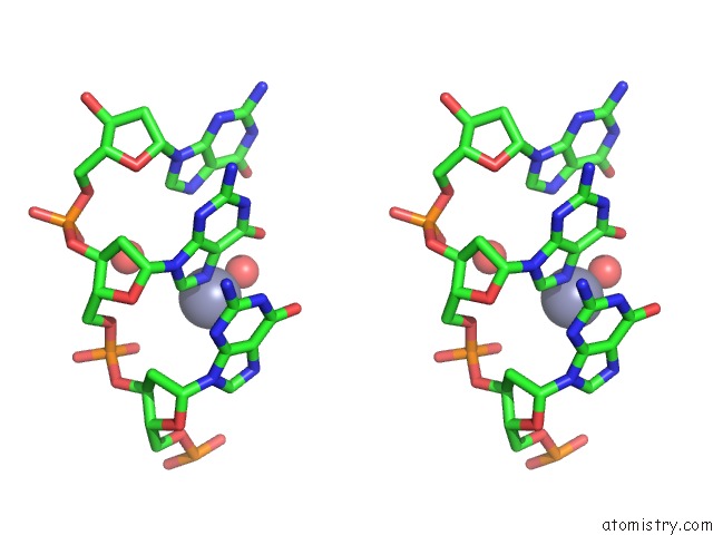
Stereo pair view
Mono view
Stereo pair view
A full contact list of Zinc with other atoms in the Zn binding
site number 1 of Comparison of X-Ray Crystal Structures of A Tetradecamer Sequence D(Cccgggtacccggg)2 at 1.7 Resolution within 5.0Å range:
|
Zinc binding site 2 out of 2 in 5iye
Go back to
Zinc binding site 2 out
of 2 in the Comparison of X-Ray Crystal Structures of A Tetradecamer Sequence D(Cccgggtacccggg)2 at 1.7 Resolution

Mono view

Stereo pair view
Mono view
Stereo pair view
A full contact list of Zinc with other atoms in the Zn binding
site number 2 of Comparison of X-Ray Crystal Structures of A Tetradecamer Sequence D(Cccgggtacccggg)2 at 1.7 Resolution within 5.0Å range:
|
Reference:
S.Karthik,
A.Thirugnanasambandam,
P.K.Mandal,
N.Gautham.
Comparison of X-Ray Crystal Structures of A Tetradecamer Sequence D(Cccgggtacccggg)2 at 1.7 Angstrom Resolution. Nucleosides Nucleotides V. 36 343 2017NUCLEIC Acids.
ISSN: ISSN 1532-2335
PubMed: 28387634
DOI: 10.1080/15257770.2017.1287378
Page generated: Sun Oct 27 18:36:34 2024
ISSN: ISSN 1532-2335
PubMed: 28387634
DOI: 10.1080/15257770.2017.1287378
Last articles
Zn in 9MJ5Zn in 9HNW
Zn in 9G0L
Zn in 9FNE
Zn in 9DZN
Zn in 9E0I
Zn in 9D32
Zn in 9DAK
Zn in 8ZXC
Zn in 8ZUF