Zinc »
PDB 5gm3-5h7r »
5h2r »
Zinc in PDB 5h2r: Crystal Structure of T Brucei Phosphodiesterase B2 Bound to Compound 15B
Protein crystallography data
The structure of Crystal Structure of T Brucei Phosphodiesterase B2 Bound to Compound 15B, PDB code: 5h2r
was solved by
C.G.Noble,
with X-Ray Crystallography technique. A brief refinement statistics is given in the table below:
| Resolution Low / High (Å) | 49.16 / 1.80 |
| Space group | P 31 2 1 |
| Cell size a, b, c (Å), α, β, γ (°) | 97.756, 97.756, 120.783, 90.00, 90.00, 120.00 |
| R / Rfree (%) | 17.6 / 22.4 |
Other elements in 5h2r:
The structure of Crystal Structure of T Brucei Phosphodiesterase B2 Bound to Compound 15B also contains other interesting chemical elements:
| Magnesium | (Mg) | 2 atoms |
Zinc Binding Sites:
The binding sites of Zinc atom in the Crystal Structure of T Brucei Phosphodiesterase B2 Bound to Compound 15B
(pdb code 5h2r). This binding sites where shown within
5.0 Angstroms radius around Zinc atom.
In total 2 binding sites of Zinc where determined in the Crystal Structure of T Brucei Phosphodiesterase B2 Bound to Compound 15B, PDB code: 5h2r:
Jump to Zinc binding site number: 1; 2;
In total 2 binding sites of Zinc where determined in the Crystal Structure of T Brucei Phosphodiesterase B2 Bound to Compound 15B, PDB code: 5h2r:
Jump to Zinc binding site number: 1; 2;
Zinc binding site 1 out of 2 in 5h2r
Go back to
Zinc binding site 1 out
of 2 in the Crystal Structure of T Brucei Phosphodiesterase B2 Bound to Compound 15B
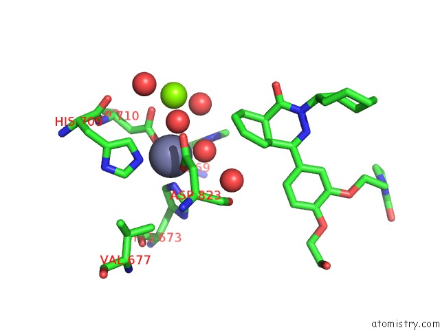
Mono view
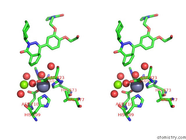
Stereo pair view
Mono view
Stereo pair view
A full contact list of Zinc with other atoms in the Zn binding
site number 1 of Crystal Structure of T Brucei Phosphodiesterase B2 Bound to Compound 15B within 5.0Å range:
|
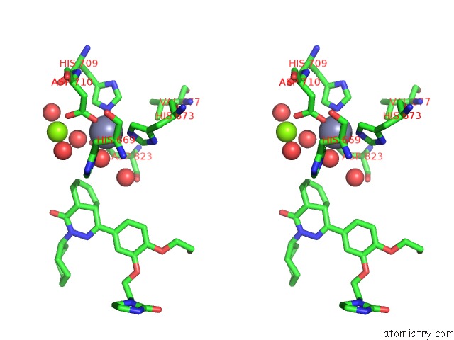
Zinc binding site 2 out of 2 in 5h2r
Go back to
Zinc binding site 2 out
of 2 in the Crystal Structure of T Brucei Phosphodiesterase B2 Bound to Compound 15B
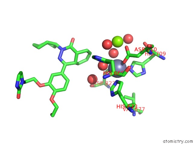
Mono view
Stereo pair view
Mono view
Stereo pair view
A full contact list of Zinc with other atoms in the Zn binding
site number 2 of Crystal Structure of T Brucei Phosphodiesterase B2 Bound to Compound 15B within 5.0Å range:
|
Reference:
P.S.Ng,
C.G.Noble,
F.S.B.Ng,
S.H.Chew,
C.C.Lim,
V.Manoharan,
K.F.Wan,
P.Vachaspati,
M.Kaiser,
N.L.Ma,
P.Gedeck,
C.Kounde,
S.P.S.Rao.
Trypanosomal Phosphodiesterase B1 and B2 As A Potential Therapy For Human African Trypanosomiasis To Be Published.
Page generated: Sun Oct 27 17:17:40 2024
Last articles
Zn in 9MJ5Zn in 9HNW
Zn in 9G0L
Zn in 9FNE
Zn in 9DZN
Zn in 9E0I
Zn in 9D32
Zn in 9DAK
Zn in 8ZXC
Zn in 8ZUF