Zinc »
PDB 5fib-5fpf »
5flx »
Zinc in PDB 5flx: Mammalian 40S Hcv-Ires Complex
Enzymatic activity of Mammalian 40S Hcv-Ires Complex
All present enzymatic activity of Mammalian 40S Hcv-Ires Complex:
4.2.99.18;
4.2.99.18;
Other elements in 5flx:
The structure of Mammalian 40S Hcv-Ires Complex also contains other interesting chemical elements:
| Magnesium | (Mg) | 74 atoms |
Zinc Binding Sites:
The binding sites of Zinc atom in the Mammalian 40S Hcv-Ires Complex
(pdb code 5flx). This binding sites where shown within
5.0 Angstroms radius around Zinc atom.
In total 2 binding sites of Zinc where determined in the Mammalian 40S Hcv-Ires Complex, PDB code: 5flx:
Jump to Zinc binding site number: 1; 2;
In total 2 binding sites of Zinc where determined in the Mammalian 40S Hcv-Ires Complex, PDB code: 5flx:
Jump to Zinc binding site number: 1; 2;
Zinc binding site 1 out of 2 in 5flx
Go back to
Zinc binding site 1 out
of 2 in the Mammalian 40S Hcv-Ires Complex
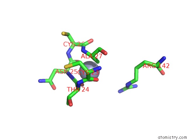
Mono view
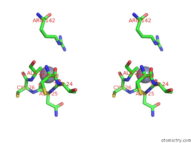
Stereo pair view
Mono view
Stereo pair view
A full contact list of Zinc with other atoms in the Zn binding
site number 1 of Mammalian 40S Hcv-Ires Complex within 5.0Å range:
|
Zinc binding site 2 out of 2 in 5flx
Go back to
Zinc binding site 2 out
of 2 in the Mammalian 40S Hcv-Ires Complex
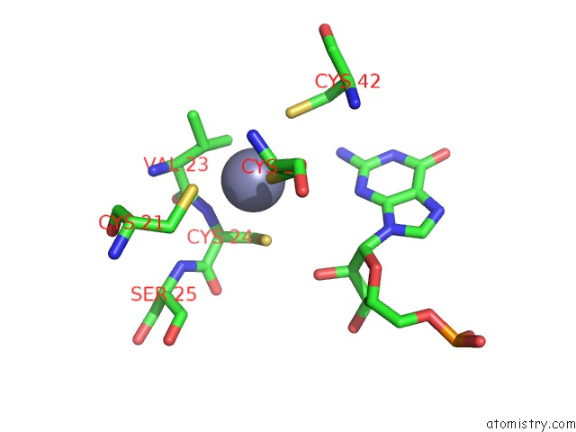
Mono view
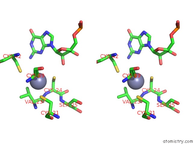
Stereo pair view
Mono view
Stereo pair view
A full contact list of Zinc with other atoms in the Zn binding
site number 2 of Mammalian 40S Hcv-Ires Complex within 5.0Å range:
|
Reference:
H.Yamamoto,
M.Collier,
J.Loerke,
J.Ismer,
A.Schmidt,
T.Hilal,
T.Sprink,
K.Yamamoto,
T.Mielke,
J.Burger,
T.R.Shaikh,
M.Dabrowski,
P.W.Hildebrand,
P.Scheerer,
C.M.Spahn.
Molecular Architecture of the Ribosome-Bound Hepatitis C Virus Internal Ribosomal Entry Site Rna. Embo J. V. 34 3042 2015.
ISSN: ISSN 0261-4189
PubMed: 26604301
DOI: 10.15252/EMBJ.201592469
Page generated: Sun Oct 27 16:20:10 2024
ISSN: ISSN 0261-4189
PubMed: 26604301
DOI: 10.15252/EMBJ.201592469
Last articles
Zn in 9MJ5Zn in 9HNW
Zn in 9G0L
Zn in 9FNE
Zn in 9DZN
Zn in 9E0I
Zn in 9D32
Zn in 9DAK
Zn in 8ZXC
Zn in 8ZUF