Zinc »
PDB 5cdt-5cvm »
5cup »
Zinc in PDB 5cup: Structure of Rhodopseudomonas Palustris Pdul - Phosphate Bound Form
Enzymatic activity of Structure of Rhodopseudomonas Palustris Pdul - Phosphate Bound Form
All present enzymatic activity of Structure of Rhodopseudomonas Palustris Pdul - Phosphate Bound Form:
2.3.1.222;
2.3.1.222;
Protein crystallography data
The structure of Structure of Rhodopseudomonas Palustris Pdul - Phosphate Bound Form, PDB code: 5cup
was solved by
M.Sutter,
O.Erbilgin,
C.A.Kerfeld,
with X-Ray Crystallography technique. A brief refinement statistics is given in the table below:
| Resolution Low / High (Å) | 39.23 / 2.10 |
| Space group | P 21 21 21 |
| Cell size a, b, c (Å), α, β, γ (°) | 57.098, 58.763, 136.656, 90.00, 90.00, 90.00 |
| R / Rfree (%) | 19.5 / 22.8 |
Zinc Binding Sites:
The binding sites of Zinc atom in the Structure of Rhodopseudomonas Palustris Pdul - Phosphate Bound Form
(pdb code 5cup). This binding sites where shown within
5.0 Angstroms radius around Zinc atom.
In total 4 binding sites of Zinc where determined in the Structure of Rhodopseudomonas Palustris Pdul - Phosphate Bound Form, PDB code: 5cup:
Jump to Zinc binding site number: 1; 2; 3; 4;
In total 4 binding sites of Zinc where determined in the Structure of Rhodopseudomonas Palustris Pdul - Phosphate Bound Form, PDB code: 5cup:
Jump to Zinc binding site number: 1; 2; 3; 4;
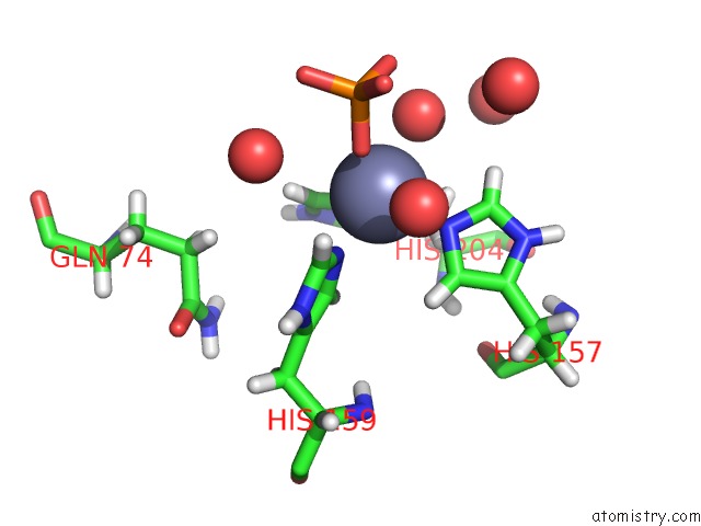
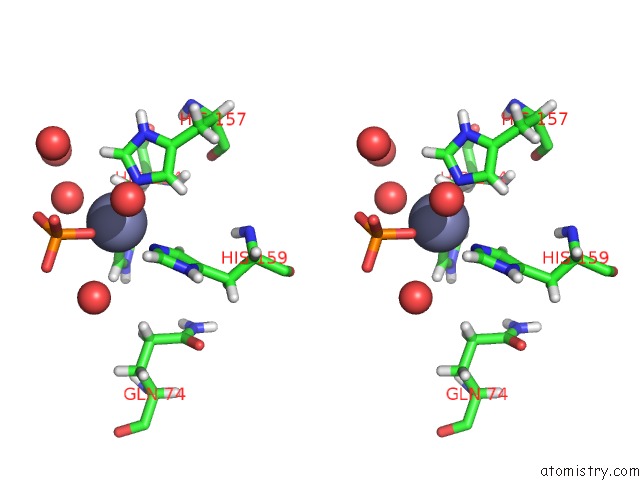
Zinc binding site 1 out of 4 in 5cup
Go back to
Zinc binding site 1 out
of 4 in the Structure of Rhodopseudomonas Palustris Pdul - Phosphate Bound Form
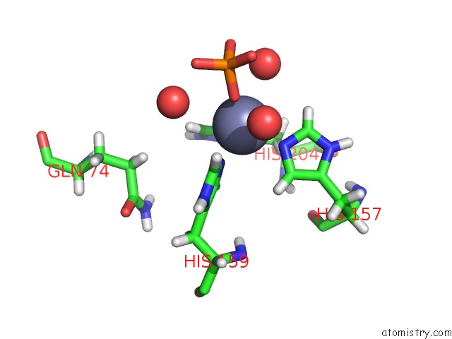
Mono view
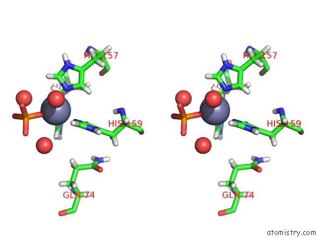
Stereo pair view
Mono view
Stereo pair view
A full contact list of Zinc with other atoms in the Zn binding
site number 1 of Structure of Rhodopseudomonas Palustris Pdul - Phosphate Bound Form within 5.0Å range:
|
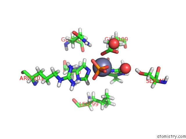
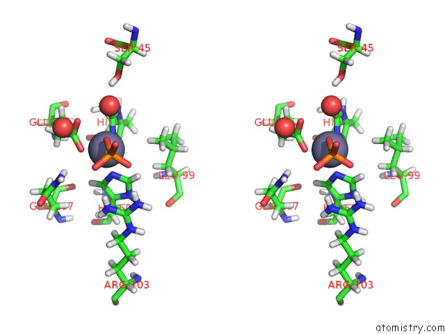
Zinc binding site 2 out of 4 in 5cup
Go back to
Zinc binding site 2 out
of 4 in the Structure of Rhodopseudomonas Palustris Pdul - Phosphate Bound Form
Mono view
Stereo pair view
Mono view
Stereo pair view
A full contact list of Zinc with other atoms in the Zn binding
site number 2 of Structure of Rhodopseudomonas Palustris Pdul - Phosphate Bound Form within 5.0Å range:
|
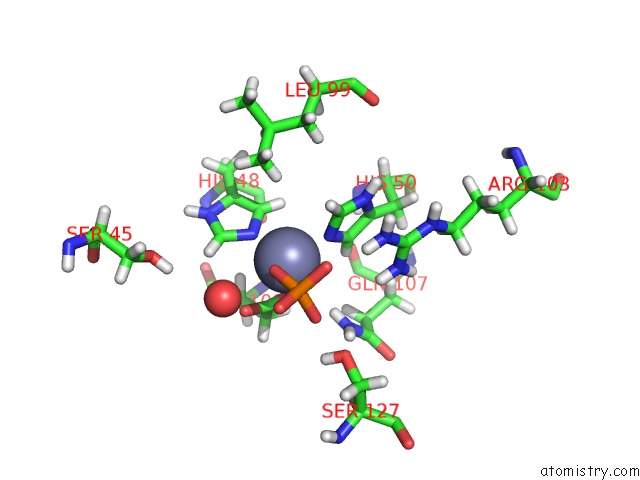
Zinc binding site 3 out of 4 in 5cup
Go back to
Zinc binding site 3 out
of 4 in the Structure of Rhodopseudomonas Palustris Pdul - Phosphate Bound Form
Mono view
Stereo pair view
Mono view
Stereo pair view
A full contact list of Zinc with other atoms in the Zn binding
site number 3 of Structure of Rhodopseudomonas Palustris Pdul - Phosphate Bound Form within 5.0Å range:
|
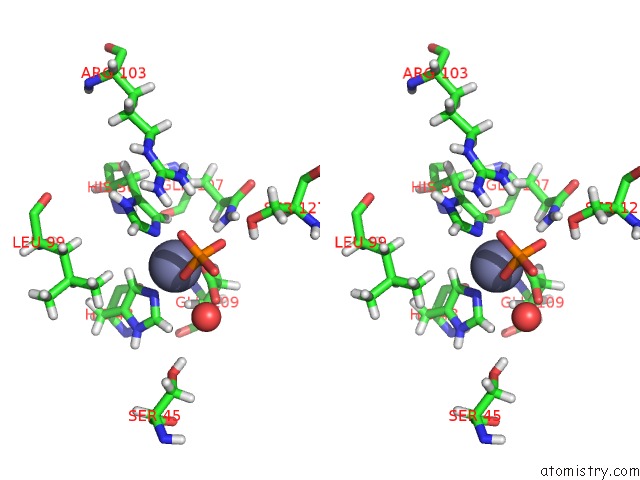
Zinc binding site 4 out of 4 in 5cup
Go back to
Zinc binding site 4 out
of 4 in the Structure of Rhodopseudomonas Palustris Pdul - Phosphate Bound Form
Mono view
Stereo pair view
Mono view
Stereo pair view
A full contact list of Zinc with other atoms in the Zn binding
site number 4 of Structure of Rhodopseudomonas Palustris Pdul - Phosphate Bound Form within 5.0Å range:
|
Reference:
O.Erbilgin,
M.Sutter,
C.A.Kerfeld.
The Structural Basis of Coenzyme A Recycling in A Bacterial Organelle. Plos Biol. V. 14 02399 2016.
ISSN: ESSN 1545-7885
PubMed: 26959993
DOI: 10.1371/JOURNAL.PBIO.1002399
Page generated: Sun Oct 27 14:28:02 2024
ISSN: ESSN 1545-7885
PubMed: 26959993
DOI: 10.1371/JOURNAL.PBIO.1002399
Last articles
Zn in 9MJ5Zn in 9HNW
Zn in 9G0L
Zn in 9FNE
Zn in 9DZN
Zn in 9E0I
Zn in 9D32
Zn in 9DAK
Zn in 8ZXC
Zn in 8ZUF