Zinc »
PDB 5cdt-5cvm »
5cjo »
Zinc in PDB 5cjo: Crystal Structure Analysis of Elbow-Engineered-Fab-Bound Human Insulin Degrading Enzyme (Ide) in Complex with Insulin
Enzymatic activity of Crystal Structure Analysis of Elbow-Engineered-Fab-Bound Human Insulin Degrading Enzyme (Ide) in Complex with Insulin
All present enzymatic activity of Crystal Structure Analysis of Elbow-Engineered-Fab-Bound Human Insulin Degrading Enzyme (Ide) in Complex with Insulin:
3.4.24.56;
3.4.24.56;
Protein crystallography data
The structure of Crystal Structure Analysis of Elbow-Engineered-Fab-Bound Human Insulin Degrading Enzyme (Ide) in Complex with Insulin, PDB code: 5cjo
was solved by
W.G.Liang,
L.Bailey,
W.J.Tang,
with X-Ray Crystallography technique. A brief refinement statistics is given in the table below:
| Resolution Low / High (Å) | 47.45 / 3.29 |
| Space group | P 2 21 21 |
| Cell size a, b, c (Å), α, β, γ (°) | 75.689, 109.218, 263.405, 90.00, 90.00, 90.00 |
| R / Rfree (%) | 19.6 / 24.5 |
Zinc Binding Sites:
The binding sites of Zinc atom in the Crystal Structure Analysis of Elbow-Engineered-Fab-Bound Human Insulin Degrading Enzyme (Ide) in Complex with Insulin
(pdb code 5cjo). This binding sites where shown within
5.0 Angstroms radius around Zinc atom.
In total only one binding site of Zinc was determined in the Crystal Structure Analysis of Elbow-Engineered-Fab-Bound Human Insulin Degrading Enzyme (Ide) in Complex with Insulin, PDB code: 5cjo:
In total only one binding site of Zinc was determined in the Crystal Structure Analysis of Elbow-Engineered-Fab-Bound Human Insulin Degrading Enzyme (Ide) in Complex with Insulin, PDB code: 5cjo:
Zinc binding site 1 out of 1 in 5cjo
Go back to
Zinc binding site 1 out
of 1 in the Crystal Structure Analysis of Elbow-Engineered-Fab-Bound Human Insulin Degrading Enzyme (Ide) in Complex with Insulin
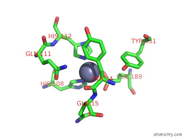
Mono view
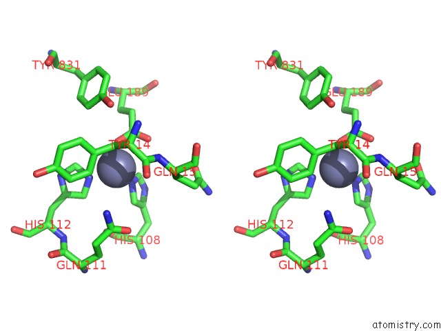
Stereo pair view
Mono view
Stereo pair view
A full contact list of Zinc with other atoms in the Zn binding
site number 1 of Crystal Structure Analysis of Elbow-Engineered-Fab-Bound Human Insulin Degrading Enzyme (Ide) in Complex with Insulin within 5.0Å range:
|
Reference:
L.J.Bailey,
K.M.Sheehy,
P.K.Dominik,
W.G.Liang,
H.Rui,
M.Clark,
M.Jaskolowski,
Y.Kim,
D.Deneka,
W.J.Tang,
A.A.Kossiakoff.
Locking the Elbow: Improved Antibody Fab Fragments As Chaperones For Structure Determination. J. Mol. Biol. 2017.
ISSN: ESSN 1089-8638
PubMed: 29273204
DOI: 10.1016/J.JMB.2017.12.012
Page generated: Sun Oct 27 14:23:57 2024
ISSN: ESSN 1089-8638
PubMed: 29273204
DOI: 10.1016/J.JMB.2017.12.012
Last articles
Zn in 9MJ5Zn in 9HNW
Zn in 9G0L
Zn in 9FNE
Zn in 9DZN
Zn in 9E0I
Zn in 9D32
Zn in 9DAK
Zn in 8ZXC
Zn in 8ZUF