Zinc »
PDB 5c4a-5cds »
5cd4 »
Zinc in PDB 5cd4: The Type Ie Crispr Cascade Complex From E. Coli, with Two Assemblies in the Asymmetric Unit Arranged Back-to-Back
Protein crystallography data
The structure of The Type Ie Crispr Cascade Complex From E. Coli, with Two Assemblies in the Asymmetric Unit Arranged Back-to-Back, PDB code: 5cd4
was solved by
R.N.Jackson,
S.M.Golden,
J.Carter,
B.Wiedenheft,
with X-Ray Crystallography technique. A brief refinement statistics is given in the table below:
| Resolution Low / High (Å) | 49.74 / 3.20 |
| Space group | P 21 21 21 |
| Cell size a, b, c (Å), α, β, γ (°) | 105.990, 244.800, 426.740, 90.00, 90.00, 90.00 |
| R / Rfree (%) | 21.2 / 25 |
Zinc Binding Sites:
The binding sites of Zinc atom in the The Type Ie Crispr Cascade Complex From E. Coli, with Two Assemblies in the Asymmetric Unit Arranged Back-to-Back
(pdb code 5cd4). This binding sites where shown within
5.0 Angstroms radius around Zinc atom.
In total 2 binding sites of Zinc where determined in the The Type Ie Crispr Cascade Complex From E. Coli, with Two Assemblies in the Asymmetric Unit Arranged Back-to-Back, PDB code: 5cd4:
Jump to Zinc binding site number: 1; 2;
In total 2 binding sites of Zinc where determined in the The Type Ie Crispr Cascade Complex From E. Coli, with Two Assemblies in the Asymmetric Unit Arranged Back-to-Back, PDB code: 5cd4:
Jump to Zinc binding site number: 1; 2;
Zinc binding site 1 out of 2 in 5cd4
Go back to
Zinc binding site 1 out
of 2 in the The Type Ie Crispr Cascade Complex From E. Coli, with Two Assemblies in the Asymmetric Unit Arranged Back-to-Back
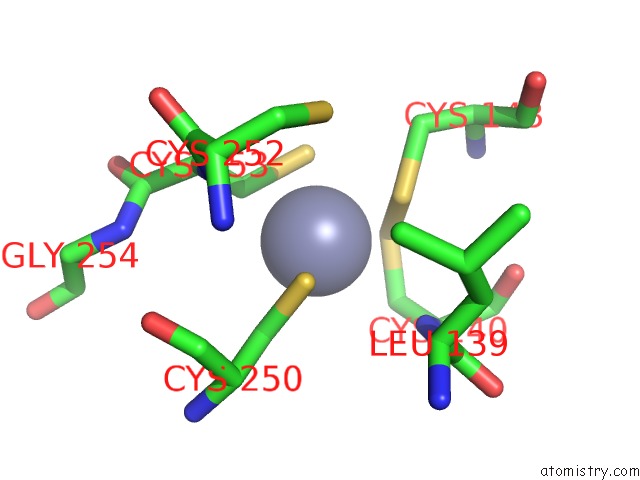
Mono view
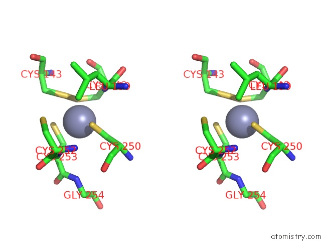
Stereo pair view
Mono view
Stereo pair view
A full contact list of Zinc with other atoms in the Zn binding
site number 1 of The Type Ie Crispr Cascade Complex From E. Coli, with Two Assemblies in the Asymmetric Unit Arranged Back-to-Back within 5.0Å range:
|
Zinc binding site 2 out of 2 in 5cd4
Go back to
Zinc binding site 2 out
of 2 in the The Type Ie Crispr Cascade Complex From E. Coli, with Two Assemblies in the Asymmetric Unit Arranged Back-to-Back
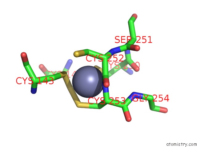
Mono view
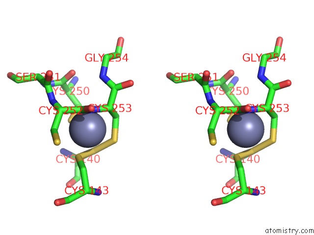
Stereo pair view
Mono view
Stereo pair view
A full contact list of Zinc with other atoms in the Zn binding
site number 2 of The Type Ie Crispr Cascade Complex From E. Coli, with Two Assemblies in the Asymmetric Unit Arranged Back-to-Back within 5.0Å range:
|
Reference:
P.B.Van Erp,
R.N.Jackson,
J.Carter,
S.M.Golden,
S.Bailey,
B.Wiedenheft.
Mechanism of Crispr-Rna Guided Recognition of Dna Targets in Escherichia Coli. Nucleic Acids Res. V. 43 8381 2015.
ISSN: ESSN 1362-4962
PubMed: 26243775
DOI: 10.1093/NAR/GKV793
Page generated: Sun Oct 27 14:11:17 2024
ISSN: ESSN 1362-4962
PubMed: 26243775
DOI: 10.1093/NAR/GKV793
Last articles
Al in 8B9IAl in 8B9G
Al in 7YLX
Al in 7YLW
Al in 7X0V
Al in 7X0S
Al in 7X24
Al in 7X22
Al in 7X21
Al in 7X20