Zinc »
PDB 5c4j-5cdt »
5c8i »
Zinc in PDB 5c8i: Joint X-Ray/Neutron Structure of Human Carbonic Anhydrase II in Complex with Methazolamide
Enzymatic activity of Joint X-Ray/Neutron Structure of Human Carbonic Anhydrase II in Complex with Methazolamide
All present enzymatic activity of Joint X-Ray/Neutron Structure of Human Carbonic Anhydrase II in Complex with Methazolamide:
4.2.1.1;
4.2.1.1;
Protein crystallography data
The structure of Joint X-Ray/Neutron Structure of Human Carbonic Anhydrase II in Complex with Methazolamide, PDB code: 5c8i
was solved by
M.Aggarwal,
A.Y.Kovalevsky,
S.Z.Fisher,
R.Mckenna,
with X-Ray Crystallography technique. A brief refinement statistics is given in the table below:
| Resolution Low / High (Å) | N/A / 1.56 |
| Space group | P 1 21 1 |
| Cell size a, b, c (Å), α, β, γ (°) | 42.893, 41.763, 72.949, 90.00, 104.59, 90.00 |
| R / Rfree (%) | 22.5 / 27.6 |
Zinc Binding Sites:
The binding sites of Zinc atom in the Joint X-Ray/Neutron Structure of Human Carbonic Anhydrase II in Complex with Methazolamide
(pdb code 5c8i). This binding sites where shown within
5.0 Angstroms radius around Zinc atom.
In total only one binding site of Zinc was determined in the Joint X-Ray/Neutron Structure of Human Carbonic Anhydrase II in Complex with Methazolamide, PDB code: 5c8i:
In total only one binding site of Zinc was determined in the Joint X-Ray/Neutron Structure of Human Carbonic Anhydrase II in Complex with Methazolamide, PDB code: 5c8i:
Zinc binding site 1 out of 1 in 5c8i
Go back to
Zinc binding site 1 out
of 1 in the Joint X-Ray/Neutron Structure of Human Carbonic Anhydrase II in Complex with Methazolamide
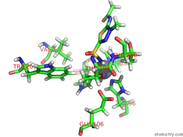
Mono view
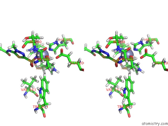
Stereo pair view
Mono view
Stereo pair view
A full contact list of Zinc with other atoms in the Zn binding
site number 1 of Joint X-Ray/Neutron Structure of Human Carbonic Anhydrase II in Complex with Methazolamide within 5.0Å range:
|
Reference:
M.Aggarwal,
A.Y.Kovalevsky,
H.Velazquez,
S.Z.Fisher,
J.C.Smith,
R.Mckenna.
Neutron Structure of Human Carbonic Anhydrase II in Complex with Methazolamide: Mapping the Solvent and Hydrogen-Bonding Patterns of An Effective Clinical Drug. Iucrj V. 3 319 2016.
ISSN: ESSN 2052-2525
PubMed: 28461893
DOI: 10.1107/S2052252516010514
Page generated: Thu Aug 21 01:14:08 2025
ISSN: ESSN 2052-2525
PubMed: 28461893
DOI: 10.1107/S2052252516010514
Last articles
Zn in 5PJIZn in 5PJG
Zn in 5PJF
Zn in 5PJH
Zn in 5PJE
Zn in 5PJD
Zn in 5PJC
Zn in 5PJB
Zn in 5PJA
Zn in 5PJ8