Zinc »
PDB 5brv-5c3k »
5bwo »
Zinc in PDB 5bwo: Crystal Structure of Human SIRT3 in Complex with A Palmitoyl H3K9 Peptide
Protein crystallography data
The structure of Crystal Structure of Human SIRT3 in Complex with A Palmitoyl H3K9 Peptide, PDB code: 5bwo
was solved by
W.Gai,
H.Jiang,
D.Liu,
with X-Ray Crystallography technique. A brief refinement statistics is given in the table below:
| Resolution Low / High (Å) | 29.15 / 2.38 |
| Space group | P 21 21 21 |
| Cell size a, b, c (Å), α, β, γ (°) | 43.445, 54.636, 113.203, 90.00, 90.00, 90.00 |
| R / Rfree (%) | 23.9 / 28.7 |
Zinc Binding Sites:
The binding sites of Zinc atom in the Crystal Structure of Human SIRT3 in Complex with A Palmitoyl H3K9 Peptide
(pdb code 5bwo). This binding sites where shown within
5.0 Angstroms radius around Zinc atom.
In total only one binding site of Zinc was determined in the Crystal Structure of Human SIRT3 in Complex with A Palmitoyl H3K9 Peptide, PDB code: 5bwo:
In total only one binding site of Zinc was determined in the Crystal Structure of Human SIRT3 in Complex with A Palmitoyl H3K9 Peptide, PDB code: 5bwo:
Zinc binding site 1 out of 1 in 5bwo
Go back to
Zinc binding site 1 out
of 1 in the Crystal Structure of Human SIRT3 in Complex with A Palmitoyl H3K9 Peptide
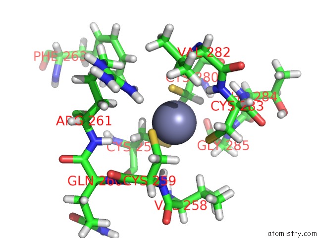
Mono view
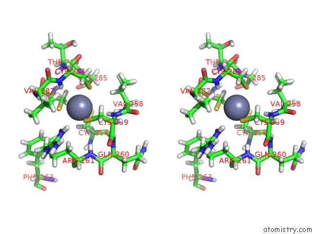
Stereo pair view
Mono view
Stereo pair view
A full contact list of Zinc with other atoms in the Zn binding
site number 1 of Crystal Structure of Human SIRT3 in Complex with A Palmitoyl H3K9 Peptide within 5.0Å range:
|
Reference:
W.Gai,
H.Li,
H.Jiang,
Y.Long,
D.Liu.
Crystal Structures of SIRT3 Reveal That the Alpha 2-Alpha 3 Loop and Alpha 3-Helix Affect the Interaction with Long-Chain Acyl Lysine. Febs Lett. V. 590 3019 2016.
ISSN: ISSN 0014-5793
PubMed: 27501476
DOI: 10.1002/1873-3468.12345
Page generated: Sun Oct 27 13:36:34 2024
ISSN: ISSN 0014-5793
PubMed: 27501476
DOI: 10.1002/1873-3468.12345
Last articles
Zn in 9MJ5Zn in 9HNW
Zn in 9G0L
Zn in 9FNE
Zn in 9DZN
Zn in 9E0I
Zn in 9D32
Zn in 9DAK
Zn in 8ZXC
Zn in 8ZUF