Zinc »
PDB 5a23-5ab9 »
5a39 »
Zinc in PDB 5a39: Structure of RAD14 in Complex with Cisplatin Containing Dna
Protein crystallography data
The structure of Structure of RAD14 in Complex with Cisplatin Containing Dna, PDB code: 5a39
was solved by
S.C.Koch,
J.Kuper,
K.L.Gasteiger,
N.Wichlein,
R.Strasser,
D.Eisen,
S.Geiger,
S.Schneider,
C.Kisker,
T.Carell,
with X-Ray Crystallography technique. A brief refinement statistics is given in the table below:
| Resolution Low / High (Å) | 54.42 / 2.80 |
| Space group | P 41 |
| Cell size a, b, c (Å), α, β, γ (°) | 54.421, 54.421, 130.773, 90.00, 90.00, 90.00 |
| R / Rfree (%) | 21.255 / 25.554 |
Other elements in 5a39:
The structure of Structure of RAD14 in Complex with Cisplatin Containing Dna also contains other interesting chemical elements:
| Platinum | (Pt) | 4 atoms |
Zinc Binding Sites:
The binding sites of Zinc atom in the Structure of RAD14 in Complex with Cisplatin Containing Dna
(pdb code 5a39). This binding sites where shown within
5.0 Angstroms radius around Zinc atom.
In total 2 binding sites of Zinc where determined in the Structure of RAD14 in Complex with Cisplatin Containing Dna, PDB code: 5a39:
Jump to Zinc binding site number: 1; 2;
In total 2 binding sites of Zinc where determined in the Structure of RAD14 in Complex with Cisplatin Containing Dna, PDB code: 5a39:
Jump to Zinc binding site number: 1; 2;
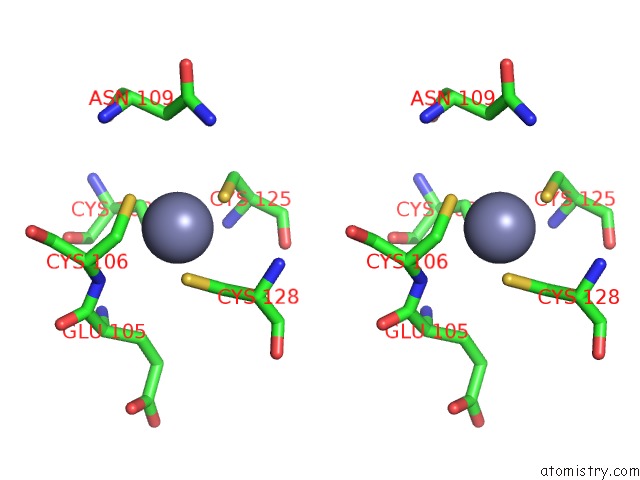
Zinc binding site 1 out of 2 in 5a39
Go back to
Zinc binding site 1 out
of 2 in the Structure of RAD14 in Complex with Cisplatin Containing Dna

Mono view
Stereo pair view
Mono view
Stereo pair view
A full contact list of Zinc with other atoms in the Zn binding
site number 1 of Structure of RAD14 in Complex with Cisplatin Containing Dna within 5.0Å range:
|
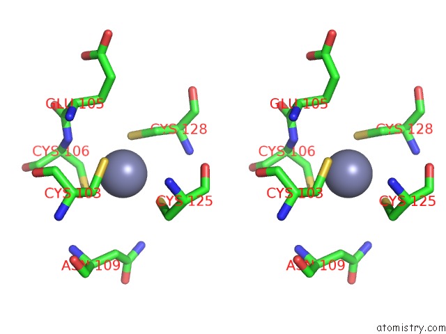
Zinc binding site 2 out of 2 in 5a39
Go back to
Zinc binding site 2 out
of 2 in the Structure of RAD14 in Complex with Cisplatin Containing Dna

Mono view
Stereo pair view
Mono view
Stereo pair view
A full contact list of Zinc with other atoms in the Zn binding
site number 2 of Structure of RAD14 in Complex with Cisplatin Containing Dna within 5.0Å range:
|
Reference:
S.C.Koch,
J.Kuper,
K.L.Gasteiger,
N.Simon,
R.Strasser,
D.Eisen,
S.Geiger,
S.Schneider,
C.Kisker,
T.Carell.
Structural Insights Into the Recognition of Cisplatin and Aaf-Dg Lesion By RAD14 (Xpa). Proc.Natl.Acad.Sci.Usa V. 112 8272 2015.
ISSN: ISSN 0027-8424
PubMed: 26100901
DOI: 10.1073/PNAS.1508509112
Page generated: Sun Oct 27 12:36:36 2024
ISSN: ISSN 0027-8424
PubMed: 26100901
DOI: 10.1073/PNAS.1508509112
Last articles
Al in 7NICAl in 7NIQ
Al in 7L07
Al in 7N77
Al in 7N73
Al in 7N72
Al in 7LVR
Al in 7KYB
Al in 7JL3
Al in 7KRO