Zinc »
PDB 4zw5-5a23 »
4zy0 »
Zinc in PDB 4zy0: X-Ray Crystal Structure of Pfa-M17 in Complex with Hydroxamic Acid- Based Inhibitor 10Q
Enzymatic activity of X-Ray Crystal Structure of Pfa-M17 in Complex with Hydroxamic Acid- Based Inhibitor 10Q
All present enzymatic activity of X-Ray Crystal Structure of Pfa-M17 in Complex with Hydroxamic Acid- Based Inhibitor 10Q:
3.4.11.1;
3.4.11.1;
Protein crystallography data
The structure of X-Ray Crystal Structure of Pfa-M17 in Complex with Hydroxamic Acid- Based Inhibitor 10Q, PDB code: 4zy0
was solved by
N.Drinkwater,
S.Mcgowan,
with X-Ray Crystallography technique. A brief refinement statistics is given in the table below:
| Resolution Low / High (Å) | 48.63 / 2.20 |
| Space group | P 21 21 21 |
| Cell size a, b, c (Å), α, β, γ (°) | 173.717, 176.072, 230.579, 90.00, 90.00, 90.00 |
| R / Rfree (%) | 19.8 / 24.4 |
Zinc Binding Sites:
Pages:
>>> Page 1 <<< Page 2, Binding sites: 11 - 20; Page 3, Binding sites: 21 - 24;Binding sites:
The binding sites of Zinc atom in the X-Ray Crystal Structure of Pfa-M17 in Complex with Hydroxamic Acid- Based Inhibitor 10Q (pdb code 4zy0). This binding sites where shown within 5.0 Angstroms radius around Zinc atom.In total 24 binding sites of Zinc where determined in the X-Ray Crystal Structure of Pfa-M17 in Complex with Hydroxamic Acid- Based Inhibitor 10Q, PDB code: 4zy0:
Jump to Zinc binding site number: 1; 2; 3; 4; 5; 6; 7; 8; 9; 10;
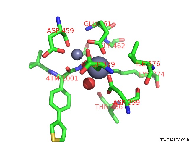
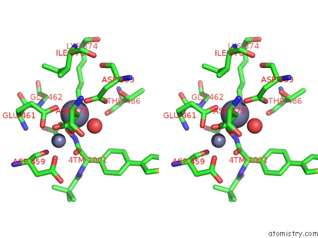
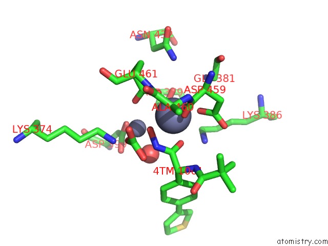
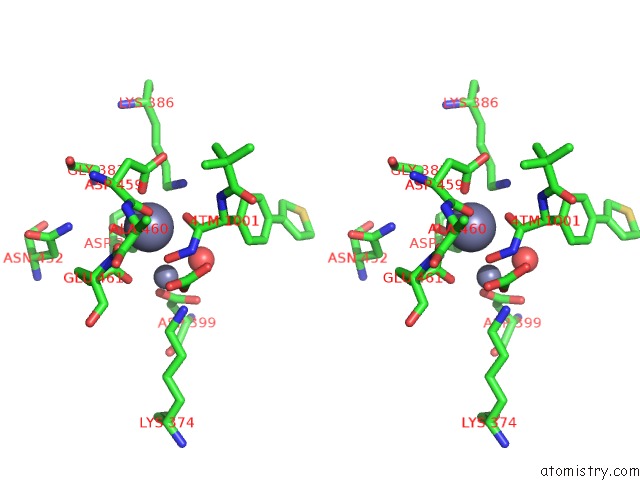
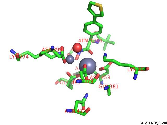
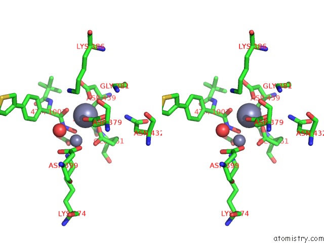
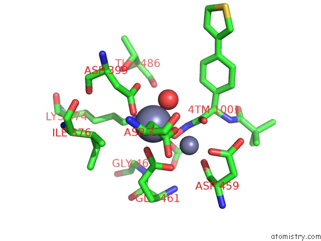
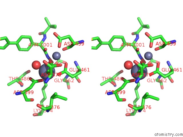
Zinc binding site 1 out of 24 in 4zy0
Go back to
Zinc binding site 1 out
of 24 in the X-Ray Crystal Structure of Pfa-M17 in Complex with Hydroxamic Acid- Based Inhibitor 10Q
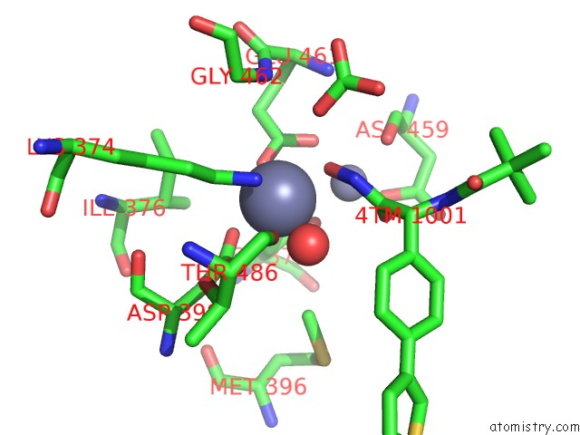
Mono view
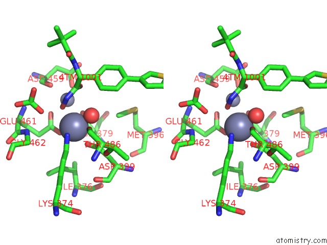
Stereo pair view
Mono view
Stereo pair view
A full contact list of Zinc with other atoms in the Zn binding
site number 1 of X-Ray Crystal Structure of Pfa-M17 in Complex with Hydroxamic Acid- Based Inhibitor 10Q within 5.0Å range:
|
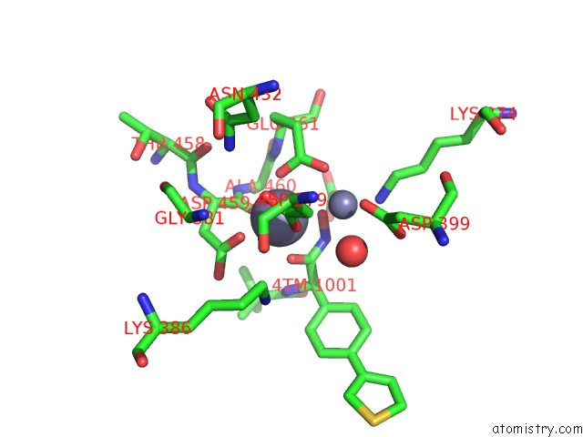
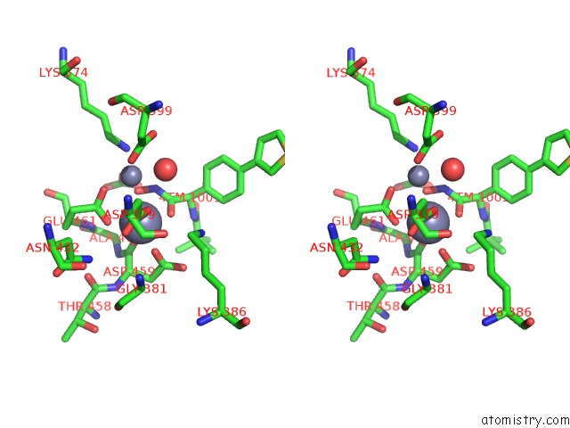
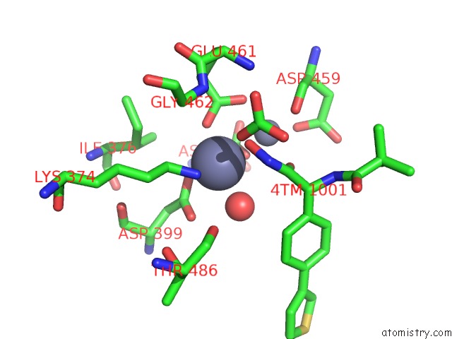
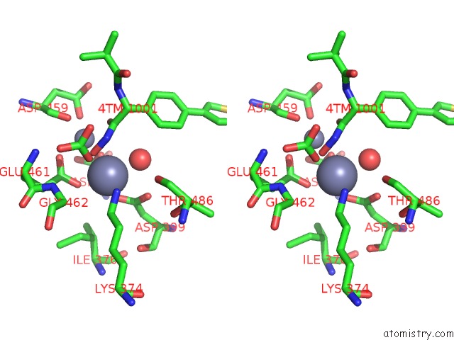
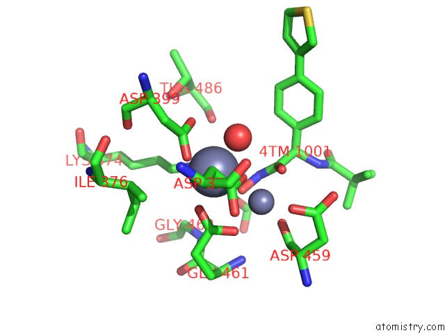
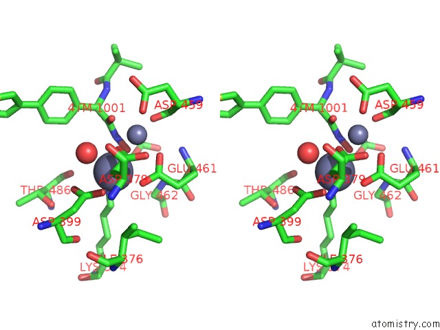
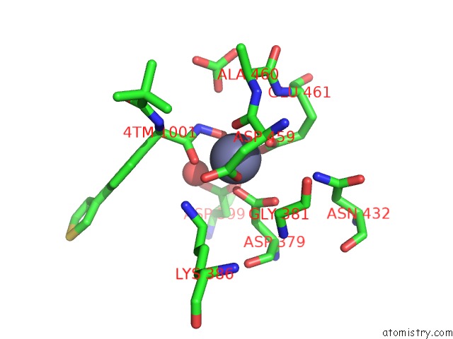
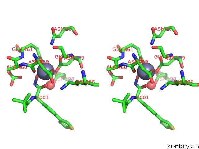
Zinc binding site 2 out of 24 in 4zy0
Go back to
Zinc binding site 2 out
of 24 in the X-Ray Crystal Structure of Pfa-M17 in Complex with Hydroxamic Acid- Based Inhibitor 10Q
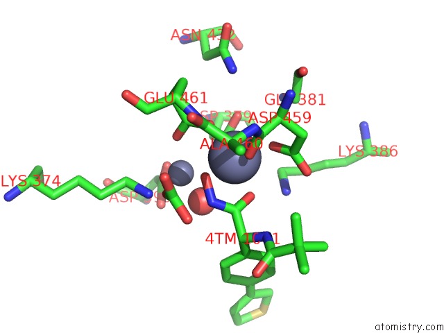
Mono view
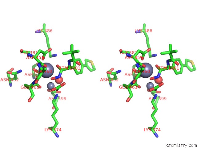
Stereo pair view
Mono view
Stereo pair view
A full contact list of Zinc with other atoms in the Zn binding
site number 2 of X-Ray Crystal Structure of Pfa-M17 in Complex with Hydroxamic Acid- Based Inhibitor 10Q within 5.0Å range:
|
Zinc binding site 3 out of 24 in 4zy0
Go back to
Zinc binding site 3 out
of 24 in the X-Ray Crystal Structure of Pfa-M17 in Complex with Hydroxamic Acid- Based Inhibitor 10Q
Mono view
Stereo pair view
Mono view
Stereo pair view
A full contact list of Zinc with other atoms in the Zn binding
site number 3 of X-Ray Crystal Structure of Pfa-M17 in Complex with Hydroxamic Acid- Based Inhibitor 10Q within 5.0Å range:
|
Zinc binding site 4 out of 24 in 4zy0
Go back to
Zinc binding site 4 out
of 24 in the X-Ray Crystal Structure of Pfa-M17 in Complex with Hydroxamic Acid- Based Inhibitor 10Q
Mono view
Stereo pair view
Mono view
Stereo pair view
A full contact list of Zinc with other atoms in the Zn binding
site number 4 of X-Ray Crystal Structure of Pfa-M17 in Complex with Hydroxamic Acid- Based Inhibitor 10Q within 5.0Å range:
|
Zinc binding site 5 out of 24 in 4zy0
Go back to
Zinc binding site 5 out
of 24 in the X-Ray Crystal Structure of Pfa-M17 in Complex with Hydroxamic Acid- Based Inhibitor 10Q
Mono view
Stereo pair view
Mono view
Stereo pair view
A full contact list of Zinc with other atoms in the Zn binding
site number 5 of X-Ray Crystal Structure of Pfa-M17 in Complex with Hydroxamic Acid- Based Inhibitor 10Q within 5.0Å range:
|
Zinc binding site 6 out of 24 in 4zy0
Go back to
Zinc binding site 6 out
of 24 in the X-Ray Crystal Structure of Pfa-M17 in Complex with Hydroxamic Acid- Based Inhibitor 10Q
Mono view
Stereo pair view
Mono view
Stereo pair view
A full contact list of Zinc with other atoms in the Zn binding
site number 6 of X-Ray Crystal Structure of Pfa-M17 in Complex with Hydroxamic Acid- Based Inhibitor 10Q within 5.0Å range:
|
Zinc binding site 7 out of 24 in 4zy0
Go back to
Zinc binding site 7 out
of 24 in the X-Ray Crystal Structure of Pfa-M17 in Complex with Hydroxamic Acid- Based Inhibitor 10Q
Mono view
Stereo pair view
Mono view
Stereo pair view
A full contact list of Zinc with other atoms in the Zn binding
site number 7 of X-Ray Crystal Structure of Pfa-M17 in Complex with Hydroxamic Acid- Based Inhibitor 10Q within 5.0Å range:
|
Zinc binding site 8 out of 24 in 4zy0
Go back to
Zinc binding site 8 out
of 24 in the X-Ray Crystal Structure of Pfa-M17 in Complex with Hydroxamic Acid- Based Inhibitor 10Q
Mono view
Stereo pair view
Mono view
Stereo pair view
A full contact list of Zinc with other atoms in the Zn binding
site number 8 of X-Ray Crystal Structure of Pfa-M17 in Complex with Hydroxamic Acid- Based Inhibitor 10Q within 5.0Å range:
|
Zinc binding site 9 out of 24 in 4zy0
Go back to
Zinc binding site 9 out
of 24 in the X-Ray Crystal Structure of Pfa-M17 in Complex with Hydroxamic Acid- Based Inhibitor 10Q
Mono view
Stereo pair view
Mono view
Stereo pair view
A full contact list of Zinc with other atoms in the Zn binding
site number 9 of X-Ray Crystal Structure of Pfa-M17 in Complex with Hydroxamic Acid- Based Inhibitor 10Q within 5.0Å range:
|
Zinc binding site 10 out of 24 in 4zy0
Go back to
Zinc binding site 10 out
of 24 in the X-Ray Crystal Structure of Pfa-M17 in Complex with Hydroxamic Acid- Based Inhibitor 10Q
Mono view
Stereo pair view
Mono view
Stereo pair view
A full contact list of Zinc with other atoms in the Zn binding
site number 10 of X-Ray Crystal Structure of Pfa-M17 in Complex with Hydroxamic Acid- Based Inhibitor 10Q within 5.0Å range:
|
Reference:
N.Drinkwater,
N.B.Vinh,
S.N.Mistry,
R.S.Bamert,
C.Ruggeri,
J.P.Holleran,
S.Loganathan,
A.Paiardini,
S.A.Charman,
A.K.Powell,
V.M.Avery,
S.Mcgowan,
P.J.Scammells.
Potent Dual Inhibitors of Plasmodium Falciparum M1 and M17 Aminopeptidases Through Optimization of S1 Pocket Interactions. Eur.J.Med.Chem. V. 110 43 2016.
ISSN: ISSN 0223-5234
PubMed: 26807544
DOI: 10.1016/J.EJMECH.2016.01.015
Page generated: Thu Aug 21 00:16:34 2025
ISSN: ISSN 0223-5234
PubMed: 26807544
DOI: 10.1016/J.EJMECH.2016.01.015
Last articles
Zn in 6G99Zn in 6G8R
Zn in 6G8V
Zn in 6G8U
Zn in 6G8B
Zn in 6G86
Zn in 6G8F
Zn in 6G7A
Zn in 6G84
Zn in 6G85