Zinc »
PDB 4yn2-4z1d »
4ywy »
Zinc in PDB 4ywy: Crystal Structure of Double Mutant Y115E Y117E Human Glutaminyl Cyclase in Complex with Inhibitor Pbd-150
Enzymatic activity of Crystal Structure of Double Mutant Y115E Y117E Human Glutaminyl Cyclase in Complex with Inhibitor Pbd-150
All present enzymatic activity of Crystal Structure of Double Mutant Y115E Y117E Human Glutaminyl Cyclase in Complex with Inhibitor Pbd-150:
2.3.2.5;
2.3.2.5;
Protein crystallography data
The structure of Crystal Structure of Double Mutant Y115E Y117E Human Glutaminyl Cyclase in Complex with Inhibitor Pbd-150, PDB code: 4ywy
was solved by
F.Di Pisa,
C.Pozzi,
M.Benvenuti,
S.Mangani,
with X-Ray Crystallography technique. A brief refinement statistics is given in the table below:
| Resolution Low / High (Å) | 32.99 / 1.95 |
| Space group | C 1 2 1 |
| Cell size a, b, c (Å), α, β, γ (°) | 86.429, 149.540, 96.210, 90.00, 96.82, 90.00 |
| R / Rfree (%) | 16.4 / 21.1 |
Other elements in 4ywy:
The structure of Crystal Structure of Double Mutant Y115E Y117E Human Glutaminyl Cyclase in Complex with Inhibitor Pbd-150 also contains other interesting chemical elements:
| Chlorine | (Cl) | 1 atom |
Zinc Binding Sites:
The binding sites of Zinc atom in the Crystal Structure of Double Mutant Y115E Y117E Human Glutaminyl Cyclase in Complex with Inhibitor Pbd-150
(pdb code 4ywy). This binding sites where shown within
5.0 Angstroms radius around Zinc atom.
In total 3 binding sites of Zinc where determined in the Crystal Structure of Double Mutant Y115E Y117E Human Glutaminyl Cyclase in Complex with Inhibitor Pbd-150, PDB code: 4ywy:
Jump to Zinc binding site number: 1; 2; 3;
In total 3 binding sites of Zinc where determined in the Crystal Structure of Double Mutant Y115E Y117E Human Glutaminyl Cyclase in Complex with Inhibitor Pbd-150, PDB code: 4ywy:
Jump to Zinc binding site number: 1; 2; 3;
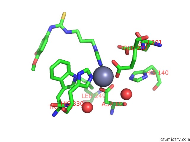
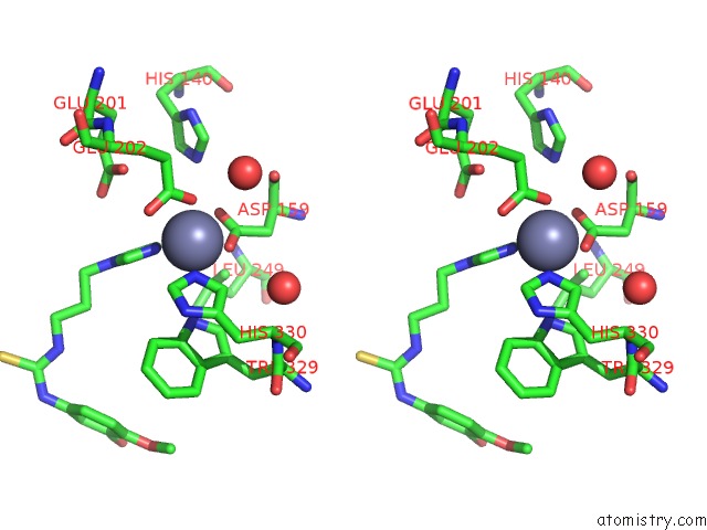
Zinc binding site 1 out of 3 in 4ywy
Go back to
Zinc binding site 1 out
of 3 in the Crystal Structure of Double Mutant Y115E Y117E Human Glutaminyl Cyclase in Complex with Inhibitor Pbd-150
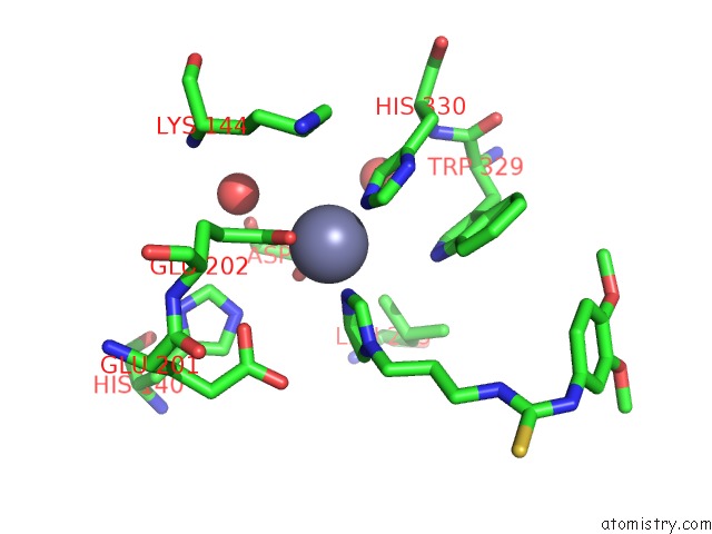
Mono view
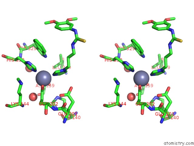
Stereo pair view
Mono view
Stereo pair view
A full contact list of Zinc with other atoms in the Zn binding
site number 1 of Crystal Structure of Double Mutant Y115E Y117E Human Glutaminyl Cyclase in Complex with Inhibitor Pbd-150 within 5.0Å range:
|
Zinc binding site 2 out of 3 in 4ywy
Go back to
Zinc binding site 2 out
of 3 in the Crystal Structure of Double Mutant Y115E Y117E Human Glutaminyl Cyclase in Complex with Inhibitor Pbd-150
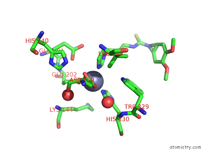
Mono view
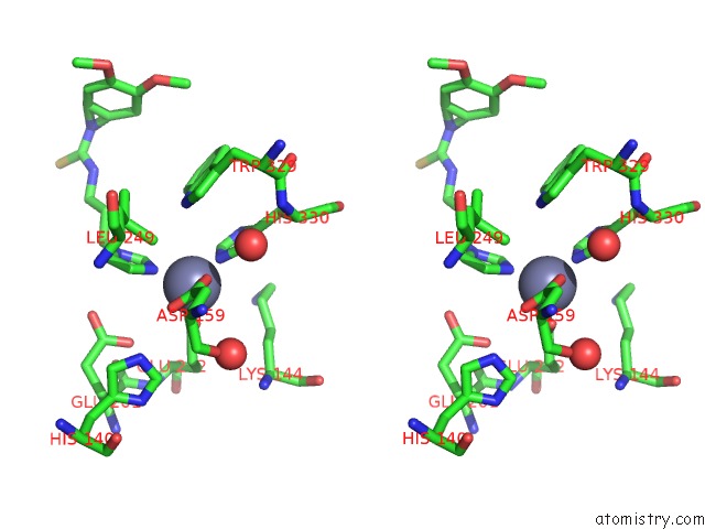
Stereo pair view
Mono view
Stereo pair view
A full contact list of Zinc with other atoms in the Zn binding
site number 2 of Crystal Structure of Double Mutant Y115E Y117E Human Glutaminyl Cyclase in Complex with Inhibitor Pbd-150 within 5.0Å range:
|
Zinc binding site 3 out of 3 in 4ywy
Go back to
Zinc binding site 3 out
of 3 in the Crystal Structure of Double Mutant Y115E Y117E Human Glutaminyl Cyclase in Complex with Inhibitor Pbd-150
Mono view
Stereo pair view
Mono view
Stereo pair view
A full contact list of Zinc with other atoms in the Zn binding
site number 3 of Crystal Structure of Double Mutant Y115E Y117E Human Glutaminyl Cyclase in Complex with Inhibitor Pbd-150 within 5.0Å range:
|
Reference:
F.Dipisa,
C.Pozzi,
M.Benvenuti,
M.Andreini,
G.Marconi,
S.Mangani.
The Soluble Y115E-Y117E Variant of Human Glutaminyl Cyclase Is A Valid Target For X-Ray and uc(Nmr) Screening of Inhibitors Against Alzheimer Disease. Acta Crystallogr.,Sect.F V. 71 986 2015.
ISSN: ESSN 2053-230X
PubMed: 26249687
DOI: 10.1107/S2053230X15010389
Page generated: Sun Oct 27 11:29:44 2024
ISSN: ESSN 2053-230X
PubMed: 26249687
DOI: 10.1107/S2053230X15010389
Last articles
Zn in 9MJ5Zn in 9HNW
Zn in 9G0L
Zn in 9FNE
Zn in 9DZN
Zn in 9E0I
Zn in 9D32
Zn in 9DAK
Zn in 8ZXC
Zn in 8ZUF