Zinc »
PDB 4xo5-4y8c »
4xsx »
Zinc in PDB 4xsx: Crystal Structure of Cbr 703 Bound to Escherichia Coli Rna Polymerase Holoenzyme
Enzymatic activity of Crystal Structure of Cbr 703 Bound to Escherichia Coli Rna Polymerase Holoenzyme
All present enzymatic activity of Crystal Structure of Cbr 703 Bound to Escherichia Coli Rna Polymerase Holoenzyme:
2.7.7.6;
2.7.7.6;
Protein crystallography data
The structure of Crystal Structure of Cbr 703 Bound to Escherichia Coli Rna Polymerase Holoenzyme, PDB code: 4xsx
was solved by
B.Bae,
S.A.Darst,
with X-Ray Crystallography technique. A brief refinement statistics is given in the table below:
| Resolution Low / High (Å) | 40.00 / 3.71 |
| Space group | P 21 21 21 |
| Cell size a, b, c (Å), α, β, γ (°) | 187.025, 206.914, 310.040, 90.00, 90.00, 90.00 |
| R / Rfree (%) | 23 / 27.3 |
Other elements in 4xsx:
The structure of Crystal Structure of Cbr 703 Bound to Escherichia Coli Rna Polymerase Holoenzyme also contains other interesting chemical elements:
| Fluorine | (F) | 6 atoms |
| Magnesium | (Mg) | 2 atoms |
Zinc Binding Sites:
The binding sites of Zinc atom in the Crystal Structure of Cbr 703 Bound to Escherichia Coli Rna Polymerase Holoenzyme
(pdb code 4xsx). This binding sites where shown within
5.0 Angstroms radius around Zinc atom.
In total 4 binding sites of Zinc where determined in the Crystal Structure of Cbr 703 Bound to Escherichia Coli Rna Polymerase Holoenzyme, PDB code: 4xsx:
Jump to Zinc binding site number: 1; 2; 3; 4;
In total 4 binding sites of Zinc where determined in the Crystal Structure of Cbr 703 Bound to Escherichia Coli Rna Polymerase Holoenzyme, PDB code: 4xsx:
Jump to Zinc binding site number: 1; 2; 3; 4;
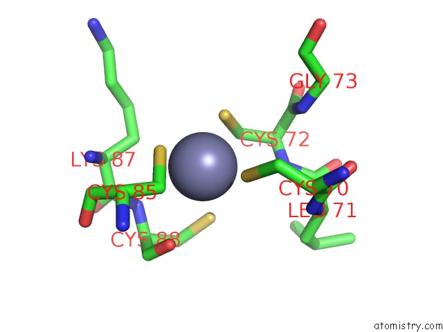
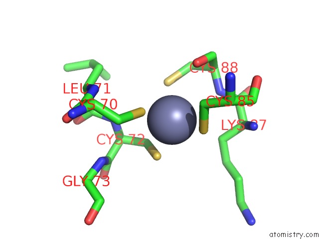
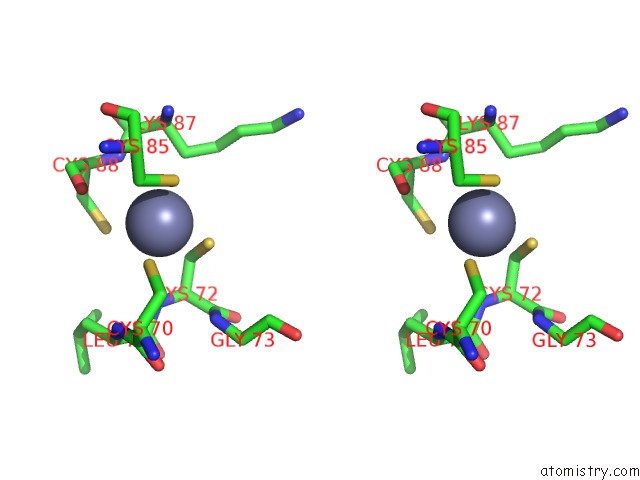
Zinc binding site 1 out of 4 in 4xsx
Go back to
Zinc binding site 1 out
of 4 in the Crystal Structure of Cbr 703 Bound to Escherichia Coli Rna Polymerase Holoenzyme
Mono view
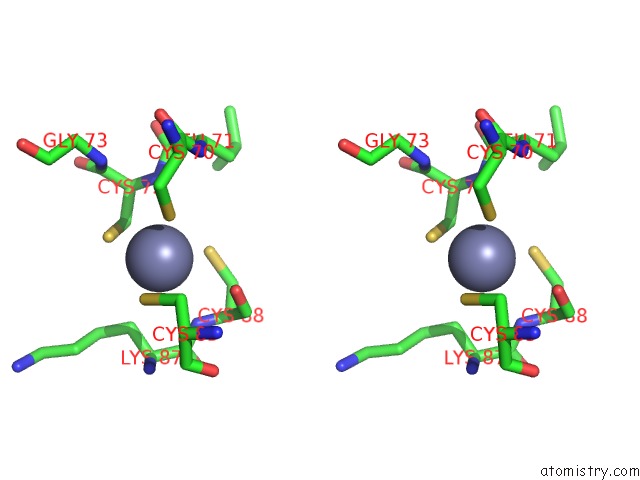
Stereo pair view
Mono view
Stereo pair view
A full contact list of Zinc with other atoms in the Zn binding
site number 1 of Crystal Structure of Cbr 703 Bound to Escherichia Coli Rna Polymerase Holoenzyme within 5.0Å range:
|
Zinc binding site 2 out of 4 in 4xsx
Go back to
Zinc binding site 2 out
of 4 in the Crystal Structure of Cbr 703 Bound to Escherichia Coli Rna Polymerase Holoenzyme
Mono view
Stereo pair view
Mono view
Stereo pair view
A full contact list of Zinc with other atoms in the Zn binding
site number 2 of Crystal Structure of Cbr 703 Bound to Escherichia Coli Rna Polymerase Holoenzyme within 5.0Å range:
|
Zinc binding site 3 out of 4 in 4xsx
Go back to
Zinc binding site 3 out
of 4 in the Crystal Structure of Cbr 703 Bound to Escherichia Coli Rna Polymerase Holoenzyme
Mono view
Stereo pair view
Mono view
Stereo pair view
A full contact list of Zinc with other atoms in the Zn binding
site number 3 of Crystal Structure of Cbr 703 Bound to Escherichia Coli Rna Polymerase Holoenzyme within 5.0Å range:
|
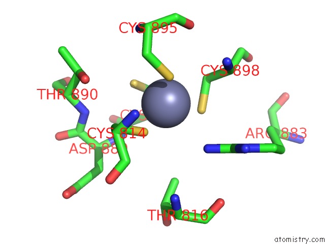
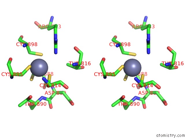
Zinc binding site 4 out of 4 in 4xsx
Go back to
Zinc binding site 4 out
of 4 in the Crystal Structure of Cbr 703 Bound to Escherichia Coli Rna Polymerase Holoenzyme
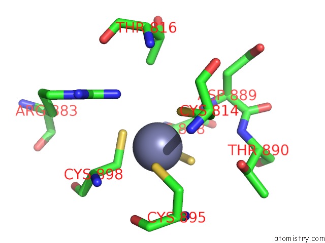
Mono view
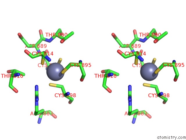
Stereo pair view
Mono view
Stereo pair view
A full contact list of Zinc with other atoms in the Zn binding
site number 4 of Crystal Structure of Cbr 703 Bound to Escherichia Coli Rna Polymerase Holoenzyme within 5.0Å range:
|
Reference:
B.Bae,
D.Nayak,
A.Ray,
A.Mustaev,
R.Landick,
S.A.Darst.
Cbr Antimicrobials Inhibit Rna Polymerase Via at Least Two Bridge-Helix Cap-Mediated Effects on Nucleotide Addition. Proc.Natl.Acad.Sci.Usa V. 112 E4178 2015.
ISSN: ESSN 1091-6490
PubMed: 26195788
DOI: 10.1073/PNAS.1502368112
Page generated: Sun Oct 27 10:46:15 2024
ISSN: ESSN 1091-6490
PubMed: 26195788
DOI: 10.1073/PNAS.1502368112
Last articles
Zn in 9MJ5Zn in 9HNW
Zn in 9G0L
Zn in 9FNE
Zn in 9DZN
Zn in 9E0I
Zn in 9D32
Zn in 9DAK
Zn in 8ZXC
Zn in 8ZUF