Atomistry » Zinc » PDB 4w6z-4wni » 4wd8
Atomistry »
  Zinc »
    PDB 4w6z-4wni »
      4wd8 »

Zinc in PDB 4wd8: Crystal Structure of A Bacterial Bestrophin Homolog From Klebsiella Pneumoniae

Protein crystallography data

The structure of Crystal Structure of A Bacterial Bestrophin Homolog From Klebsiella Pneumoniae, PDB code: 4wd8 was solved by T.Yang, Q.Liu, W.A.Hendrickson, New York Consortium On Membrane Proteinstructure (Nycomps), with X-Ray Crystallography technique. A brief refinement statistics is given in the table below:

Resolution Low / High (Å) 38.96 / 2.30
Space group P 21 21 21
Cell size a, b, c (Å), α, β, γ (°) 114.079, 160.034, 161.881, 90.00, 90.00, 90.00
R / Rfree (%) 19.6 / 21.5

Zinc Binding Sites:

Pages:

>>> Page 1 <<< Page 2, Binding sites: 11 - 15;

Binding sites:

The binding sites of Zinc atom in the Crystal Structure of A Bacterial Bestrophin Homolog From Klebsiella Pneumoniae (pdb code 4wd8). This binding sites where shown within 5.0 Angstroms radius around Zinc atom.
In total 15 binding sites of Zinc where determined in the Crystal Structure of A Bacterial Bestrophin Homolog From Klebsiella Pneumoniae, PDB code: 4wd8:
Jump to Zinc binding site number: 1; 2; 3; 4; 5; 6; 7; 8; 9; 10;

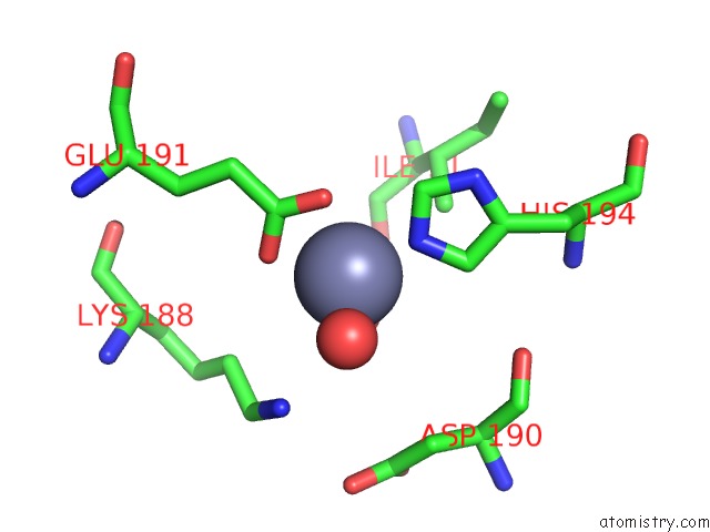
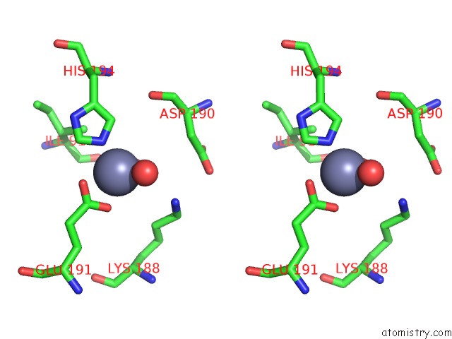
Zinc binding site 1 out of 15 in 4wd8

Go back to Zinc Binding Sites List in 4wd8
Zinc binding site 1 out of 15 in the Crystal Structure of A Bacterial Bestrophin Homolog From Klebsiella Pneumoniae


Mono view


Stereo pair view

A full contact list of Zinc with other atoms in the Zn binding site number 1 of Crystal Structure of A Bacterial Bestrophin Homolog From Klebsiella Pneumoniae within 5.0Å range:
probe atom residue distance (Å) B Occ
A:Zn301

b:34.2
occ:1.00
NE2 E:HIS194 2.0 26.1 1.0
OE2 A:GLU191 2.1 84.0 1.0
O A:HOH487 2.1 37.5 1.0
O A:HOH474 2.1 5.8 1.0
OE1 A:GLU191 2.2 67.9 1.0
CD A:GLU191 2.4 74.3 1.0
CE1 E:HIS194 3.0 23.3 1.0
CD2 E:HIS194 3.0 18.6 1.0
CG A:GLU191 4.0 45.1 1.0
ND1 E:HIS194 4.1 23.0 1.0
CG E:HIS194 4.1 23.2 1.0
CE A:LYS188 4.4 43.4 1.0
CD A:LYS188 4.5 35.3 1.0
CG2 A:ILE91 4.5 21.8 1.0
CB A:GLU191 4.9 18.9 1.0
CB E:ASP190 5.0 30.8 1.0

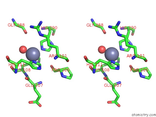
Zinc binding site 2 out of 15 in 4wd8

Go back to Zinc Binding Sites List in 4wd8
Zinc binding site 2 out of 15 in the Crystal Structure of A Bacterial Bestrophin Homolog From Klebsiella Pneumoniae


Mono view


Stereo pair view

A full contact list of Zinc with other atoms in the Zn binding site number 2 of Crystal Structure of A Bacterial Bestrophin Homolog From Klebsiella Pneumoniae within 5.0Å range:
probe atom residue distance (Å) B Occ
A:Zn302

b:37.9
occ:1.00
O A:HOH493 2.1 30.6 1.0
NE2 A:HIS194 2.1 26.4 1.0
OE2 B:GLU191 2.1 49.0 1.0
O B:HOH434 2.2 15.6 1.0
OE1 B:GLU191 2.2 42.1 1.0
CD B:GLU191 2.5 41.7 1.0
CE1 A:HIS194 3.0 28.6 1.0
CD2 A:HIS194 3.2 21.9 1.0
CG B:GLU191 4.1 42.6 1.0
ND1 A:HIS194 4.2 26.6 1.0
CE B:LYS188 4.2 41.6 1.0
CG A:HIS194 4.3 21.2 1.0
CD B:LYS188 4.3 38.9 1.0
CG2 B:ILE91 4.4 17.6 1.0
NZ B:LYS188 4.8 47.2 1.0
CB B:GLU191 5.0 21.7 1.0

Zinc binding site 3 out of 15 in 4wd8

Go back to Zinc Binding Sites List in 4wd8
Zinc binding site 3 out of 15 in the Crystal Structure of A Bacterial Bestrophin Homolog From Klebsiella Pneumoniae


Mono view


Stereo pair view

A full contact list of Zinc with other atoms in the Zn binding site number 3 of Crystal Structure of A Bacterial Bestrophin Homolog From Klebsiella Pneumoniae within 5.0Å range:
probe atom residue distance (Å) B Occ
A:Zn303

b:45.2
occ:1.00
ND1 A:HIS108 2.0 72.4 1.0
O A:HOH460 2.1 28.4 1.0
ND1 A:HIS111 2.1 36.9 1.0
CE1 A:HIS108 3.0 76.1 1.0
CE1 A:HIS111 3.0 40.6 1.0
CG A:HIS108 3.1 67.6 1.0
CG A:HIS111 3.1 38.4 1.0
CB A:HIS108 3.5 58.7 1.0
CB A:HIS111 3.5 31.3 1.0
CA A:HIS108 3.6 52.0 1.0
NE2 A:HIS108 4.1 76.4 1.0
NE2 A:HIS111 4.1 33.2 1.0
CD2 A:HIS108 4.2 72.0 1.0
CD2 A:HIS111 4.2 38.1 1.0
N A:HIS108 4.4 51.4 1.0
O A:HIS108 4.5 45.9 1.0
C A:HIS108 4.6 48.3 1.0
O A:PRO105 4.9 51.6 1.0
CB A:THR287 5.0 55.3 1.0
CG A:ARG101 5.0 44.5 1.0
CG2 A:THR287 5.0 49.6 1.0
C A:GLU107 5.0 47.9 1.0
O A:THR287 5.0 65.0 1.0

Zinc binding site 4 out of 15 in 4wd8

Go back to Zinc Binding Sites List in 4wd8
Zinc binding site 4 out of 15 in the Crystal Structure of A Bacterial Bestrophin Homolog From Klebsiella Pneumoniae


Mono view


Stereo pair view

A full contact list of Zinc with other atoms in the Zn binding site number 4 of Crystal Structure of A Bacterial Bestrophin Homolog From Klebsiella Pneumoniae within 5.0Å range:
probe atom residue distance (Å) B Occ
A:Zn304

b:43.4
occ:1.00
OD2 A:ASP269 2.0 33.8 1.0
OD2 A:ASP261 2.0 37.4 1.0
O A:HOH472 2.2 47.4 1.0
CG A:ASP269 2.9 33.1 1.0
CG A:ASP261 3.0 39.6 1.0
OD1 A:ASP269 3.2 34.5 1.0
CB A:ASP261 3.4 34.6 1.0
CB A:ALA266 3.7 31.7 1.0
CA A:GLY264 3.8 25.6 1.0
O A:ASP261 3.8 27.8 1.0
N A:ALA266 4.0 38.7 1.0
OD1 A:ASP261 4.1 39.8 1.0
N A:THR265 4.2 35.2 1.0
C A:GLY264 4.3 33.2 1.0
CB A:ASP269 4.3 31.8 1.0
O A:HOH408 4.4 47.5 1.0
CA A:ALA266 4.5 39.0 1.0
N A:GLY264 4.5 27.7 1.0
CA A:ASP261 4.7 32.5 1.0
C A:ASP261 4.7 30.8 1.0

Zinc binding site 5 out of 15 in 4wd8

Go back to Zinc Binding Sites List in 4wd8
Zinc binding site 5 out of 15 in the Crystal Structure of A Bacterial Bestrophin Homolog From Klebsiella Pneumoniae


Mono view


Stereo pair view

A full contact list of Zinc with other atoms in the Zn binding site number 5 of Crystal Structure of A Bacterial Bestrophin Homolog From Klebsiella Pneumoniae within 5.0Å range:
probe atom residue distance (Å) B Occ
B:Zn301

b:81.5
occ:1.00
ND1 B:HIS235 2.1 82.2 1.0
O B:HOH405 2.1 36.7 1.0
CG B:HIS235 2.9 79.3 1.0
CE1 B:HIS235 3.0 79.4 1.0
CB B:HIS235 3.2 76.0 1.0
CA B:HIS235 3.9 74.7 1.0
CD2 B:HIS235 4.0 79.1 1.0
NE2 B:HIS235 4.1 78.9 1.0
CD2 B:TYR42 4.4 73.3 1.0
CE2 B:TYR42 4.7 72.2 1.0
C B:HIS235 4.8 73.6 1.0
O B:VAL231 4.9 73.7 1.0

Zinc binding site 6 out of 15 in 4wd8

Go back to Zinc Binding Sites List in 4wd8
Zinc binding site 6 out of 15 in the Crystal Structure of A Bacterial Bestrophin Homolog From Klebsiella Pneumoniae


Mono view


Stereo pair view

A full contact list of Zinc with other atoms in the Zn binding site number 6 of Crystal Structure of A Bacterial Bestrophin Homolog From Klebsiella Pneumoniae within 5.0Å range:
probe atom residue distance (Å) B Occ
B:Zn302

b:38.3
occ:1.00
NE2 B:HIS194 2.0 24.8 1.0
O B:HOH441 2.1 34.1 1.0
OE2 D:GLU191 2.1 71.2 1.0
O D:HOH421 2.2 15.4 1.0
OE1 D:GLU191 2.2 67.1 1.0
CD D:GLU191 2.5 67.6 1.0
CE1 B:HIS194 3.0 26.2 1.0
CD2 B:HIS194 3.1 23.9 1.0
NZ D:LYS188 3.9 55.1 1.0
CG D:GLU191 4.1 75.2 1.0
ND1 B:HIS194 4.1 30.3 1.0
CG B:HIS194 4.2 28.0 1.0
CG2 D:ILE91 4.5 25.4 1.0
CD D:LYS188 4.7 39.1 1.0
CE D:LYS188 4.8 49.2 1.0
CB D:GLU191 4.9 23.8 1.0
CB B:ASP190 5.0 36.7 1.0

Zinc binding site 7 out of 15 in 4wd8

Go back to Zinc Binding Sites List in 4wd8
Zinc binding site 7 out of 15 in the Crystal Structure of A Bacterial Bestrophin Homolog From Klebsiella Pneumoniae


Mono view


Stereo pair view

A full contact list of Zinc with other atoms in the Zn binding site number 7 of Crystal Structure of A Bacterial Bestrophin Homolog From Klebsiella Pneumoniae within 5.0Å range:
probe atom residue distance (Å) B Occ
B:Zn303

b:40.2
occ:1.00
ND1 B:HIS111 2.1 35.2 1.0
NE2 B:HIS290 2.1 41.2 1.0
O B:HOH439 2.1 31.9 1.0
ND1 B:HIS108 2.1 47.1 1.0
CD2 B:HIS290 2.9 38.4 1.0
CE1 B:HIS111 2.9 37.8 1.0
CE1 B:HIS108 2.9 41.3 1.0
CE1 B:HIS290 3.2 47.1 1.0
CG B:HIS111 3.2 35.4 1.0
CG B:HIS108 3.2 44.7 1.0
CB B:HIS111 3.6 36.2 1.0
CB B:HIS108 3.7 41.2 1.0
CA B:HIS108 3.7 41.3 1.0
NE2 B:HIS111 4.1 35.2 1.0
CG B:HIS290 4.1 42.4 1.0
NE2 B:HIS108 4.1 43.2 1.0
ND1 B:HIS290 4.2 46.2 1.0
CD2 B:HIS111 4.2 33.7 1.0
CD2 B:HIS108 4.3 44.9 1.0
O B:GLY288 4.4 47.3 1.0
N B:HIS108 4.4 41.4 1.0
O B:PRO105 4.4 46.1 1.0
O B:HIS108 4.7 43.8 1.0
C B:HIS108 4.8 43.1 1.0
CG B:ARG101 4.8 35.4 1.0
C B:GLU107 4.9 43.8 1.0
O B:GLU107 4.9 47.8 1.0
NE B:ARG101 4.9 45.1 1.0

Zinc binding site 8 out of 15 in 4wd8

Go back to Zinc Binding Sites List in 4wd8
Zinc binding site 8 out of 15 in the Crystal Structure of A Bacterial Bestrophin Homolog From Klebsiella Pneumoniae


Mono view


Stereo pair view

A full contact list of Zinc with other atoms in the Zn binding site number 8 of Crystal Structure of A Bacterial Bestrophin Homolog From Klebsiella Pneumoniae within 5.0Å range:
probe atom residue distance (Å) B Occ
C:Zn301

b:31.6
occ:1.00
NE2 D:HIS194 2.0 29.1 1.0
O D:HOH430 2.1 28.8 1.0
OE2 C:GLU191 2.1 48.2 1.0
O C:HOH467 2.2 7.5 1.0
OE1 C:GLU191 2.2 41.2 1.0
CD C:GLU191 2.5 40.7 1.0
CE1 D:HIS194 3.0 29.3 1.0
CD2 D:HIS194 3.1 29.3 1.0
NZ C:LYS188 3.9 51.6 1.0
CG C:GLU191 4.0 35.5 1.0
ND1 D:HIS194 4.1 32.3 1.0
CG D:HIS194 4.2 29.6 1.0
O C:HOH480 4.3 40.0 1.0
CG2 C:ILE91 4.5 21.9 1.0
CD C:LYS188 4.8 40.4 1.0
CE C:LYS188 4.9 45.3 1.0
CB C:GLU191 4.9 18.4 1.0
CB D:ASP190 5.0 37.7 1.0

Zinc binding site 9 out of 15 in 4wd8

Go back to Zinc Binding Sites List in 4wd8
Zinc binding site 9 out of 15 in the Crystal Structure of A Bacterial Bestrophin Homolog From Klebsiella Pneumoniae


Mono view


Stereo pair view

A full contact list of Zinc with other atoms in the Zn binding site number 9 of Crystal Structure of A Bacterial Bestrophin Homolog From Klebsiella Pneumoniae within 5.0Å range:
probe atom residue distance (Å) B Occ
C:Zn302

b:43.0
occ:1.00
NE2 C:HIS194 2.1 22.2 1.0
OE2 E:GLU191 2.1 77.1 1.0
O E:HOH420 2.1 45.2 1.0
O C:HOH468 2.2 26.6 1.0
OE1 E:GLU191 2.2 68.1 1.0
OD1 C:ASP190 2.3 83.2 1.0
CD E:GLU191 2.5 70.3 1.0
OD2 C:ASP190 2.8 87.3 1.0
CG C:ASP190 3.0 84.5 1.0
CD2 C:HIS194 3.0 17.4 1.0
CE1 C:HIS194 3.1 22.0 1.0
O E:HOH433 4.0 54.3 1.0
CE E:LYS188 4.1 45.6 1.0
CG E:GLU191 4.1 28.8 1.0
ND1 C:HIS194 4.2 22.2 1.0
CG C:HIS194 4.2 20.1 1.0
CD E:LYS188 4.4 39.1 1.0
CG2 E:ILE91 4.5 14.1 1.0
CB C:ASP190 4.5 26.6 1.0
NZ E:LYS188 4.7 55.7 1.0
O C:ASP190 4.9 26.3 1.0

Zinc binding site 10 out of 15 in 4wd8

Go back to Zinc Binding Sites List in 4wd8
Zinc binding site 10 out of 15 in the Crystal Structure of A Bacterial Bestrophin Homolog From Klebsiella Pneumoniae


Mono view


Stereo pair view

A full contact list of Zinc with other atoms in the Zn binding site number 10 of Crystal Structure of A Bacterial Bestrophin Homolog From Klebsiella Pneumoniae within 5.0Å range:
probe atom residue distance (Å) B Occ
C:Zn303

b:38.1
occ:1.00
ND1 C:HIS111 2.1 40.8 1.0
ND1 C:HIS108 2.1 38.2 1.0
O C:HOH470 2.1 27.0 1.0
O C:HOH408 2.1 35.5 1.0
CE1 C:HIS108 2.9 38.9 1.0
CE1 C:HIS111 3.0 33.5 1.0
CG C:HIS111 3.1 31.3 1.0
CG C:HIS108 3.1 42.8 1.0
CB C:HIS111 3.4 30.9 1.0
CB C:HIS108 3.5 40.0 1.0
CA C:HIS108 3.6 41.3 1.0
NE2 C:HIS108 4.1 41.0 1.0
NE2 C:HIS111 4.1 32.7 1.0
CD2 C:HIS108 4.2 41.7 1.0
CD2 C:HIS111 4.2 31.2 1.0
N C:HIS108 4.5 40.6 1.0
O C:HIS108 4.5 42.8 1.0
C C:HIS108 4.6 40.2 1.0
CG C:ARG101 4.8 38.5 1.0
O C:PRO105 4.8 53.3 1.0
NE C:ARG101 4.8 52.5 1.0
CA C:HIS111 4.9 38.6 1.0
CD C:ARG101 5.0 42.8 1.0
O C:GLU107 5.0 38.2 1.0
C C:GLU107 5.0 38.8 1.0

Reference:

T.Yang, Q.Liu, B.Kloss, R.Bruni, R.C.Kalathur, Y.Guo, E.Kloppmann, B.Rost, H.M.Colecraft, W.A.Hendrickson. Structure and Selectivity in Bestrophin Ion Channels. Science V. 346 355 2014.
ISSN: ESSN 1095-9203
PubMed: 25324390
DOI: 10.1126/SCIENCE.1259723
Page generated: Wed Dec 16 05:50:56 2020

Last articles

Zn in 8WB0
Zn in 8WAX
Zn in 8WAU
Zn in 8WAZ
Zn in 8WAY
Zn in 8WAV
Zn in 8WAW
Zn in 8WAT
Zn in 8W7M
Zn in 8WD3
© Copyright 2008-2020 by atomistry.com
Home   |    Site Map   |    Copyright   |    Contact us   |    Privacy