Zinc »
PDB 4w8y-4wnu »
4w8y »
Zinc in PDB 4w8y: Structure of Full Length CMR2 From Pyrococcus Furiosus (Manganese Bound Form)
Protein crystallography data
The structure of Structure of Full Length CMR2 From Pyrococcus Furiosus (Manganese Bound Form), PDB code: 4w8y
was solved by
C.Benda,
J.Ebert,
M.Baumgaertner,
E.Conti,
with X-Ray Crystallography technique. A brief refinement statistics is given in the table below:
| Resolution Low / High (Å) | 85.72 / 3.00 |
| Space group | P 1 21 1 |
| Cell size a, b, c (Å), α, β, γ (°) | 62.400, 167.925, 100.908, 90.00, 98.91, 90.00 |
| R / Rfree (%) | 22.3 / 25.9 |
Other elements in 4w8y:
The structure of Structure of Full Length CMR2 From Pyrococcus Furiosus (Manganese Bound Form) also contains other interesting chemical elements:
| Manganese | (Mn) | 8 atoms |
Zinc Binding Sites:
The binding sites of Zinc atom in the Structure of Full Length CMR2 From Pyrococcus Furiosus (Manganese Bound Form)
(pdb code 4w8y). This binding sites where shown within
5.0 Angstroms radius around Zinc atom.
In total 2 binding sites of Zinc where determined in the Structure of Full Length CMR2 From Pyrococcus Furiosus (Manganese Bound Form), PDB code: 4w8y:
Jump to Zinc binding site number: 1; 2;
In total 2 binding sites of Zinc where determined in the Structure of Full Length CMR2 From Pyrococcus Furiosus (Manganese Bound Form), PDB code: 4w8y:
Jump to Zinc binding site number: 1; 2;
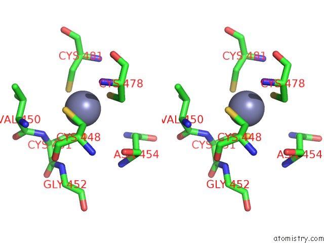
Zinc binding site 1 out of 2 in 4w8y
Go back to
Zinc binding site 1 out
of 2 in the Structure of Full Length CMR2 From Pyrococcus Furiosus (Manganese Bound Form)

Mono view
Stereo pair view
Mono view
Stereo pair view
A full contact list of Zinc with other atoms in the Zn binding
site number 1 of Structure of Full Length CMR2 From Pyrococcus Furiosus (Manganese Bound Form) within 5.0Å range:
|
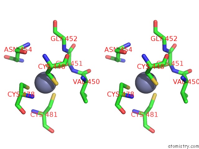
Zinc binding site 2 out of 2 in 4w8y
Go back to
Zinc binding site 2 out
of 2 in the Structure of Full Length CMR2 From Pyrococcus Furiosus (Manganese Bound Form)

Mono view
Stereo pair view
Mono view
Stereo pair view
A full contact list of Zinc with other atoms in the Zn binding
site number 2 of Structure of Full Length CMR2 From Pyrococcus Furiosus (Manganese Bound Form) within 5.0Å range:
|
Reference:
C.Benda,
J.Ebert,
R.A.Scheltema,
H.B.Schiller,
M.Baumgartner,
F.Bonneau,
M.Mann,
E.Conti.
Structural Model of A Crispr Rna-Silencing Complex Reveals the Rna-Target Cleavage Activity in CMR4. Mol.Cell V. 56 43 2014.
ISSN: ISSN 1097-2765
PubMed: 25280103
DOI: 10.1016/J.MOLCEL.2014.09.002
Page generated: Sun Oct 27 09:43:50 2024
ISSN: ISSN 1097-2765
PubMed: 25280103
DOI: 10.1016/J.MOLCEL.2014.09.002
Last articles
Zn in 9MJ5Zn in 9HNW
Zn in 9G0L
Zn in 9FNE
Zn in 9DZN
Zn in 9E0I
Zn in 9D32
Zn in 9DAK
Zn in 8ZXC
Zn in 8ZUF