Zinc »
PDB 4rvo-4tpj »
4tos »
Zinc in PDB 4tos: Crystal Structure of Tankyrase 1 with 355
Enzymatic activity of Crystal Structure of Tankyrase 1 with 355
All present enzymatic activity of Crystal Structure of Tankyrase 1 with 355:
2.4.2.30;
2.4.2.30;
Protein crystallography data
The structure of Crystal Structure of Tankyrase 1 with 355, PDB code: 4tos
was solved by
H.Chen,
X.Zhang,
L.Lum,
C.Chen,
with X-Ray Crystallography technique. A brief refinement statistics is given in the table below:
| Resolution Low / High (Å) | 31.00 / 1.80 |
| Space group | P 21 21 21 |
| Cell size a, b, c (Å), α, β, γ (°) | 48.195, 81.168, 114.155, 90.00, 90.00, 90.00 |
| R / Rfree (%) | 18.1 / 21.1 |
Zinc Binding Sites:
The binding sites of Zinc atom in the Crystal Structure of Tankyrase 1 with 355
(pdb code 4tos). This binding sites where shown within
5.0 Angstroms radius around Zinc atom.
In total 2 binding sites of Zinc where determined in the Crystal Structure of Tankyrase 1 with 355, PDB code: 4tos:
Jump to Zinc binding site number: 1; 2;
In total 2 binding sites of Zinc where determined in the Crystal Structure of Tankyrase 1 with 355, PDB code: 4tos:
Jump to Zinc binding site number: 1; 2;
Zinc binding site 1 out of 2 in 4tos
Go back to
Zinc binding site 1 out
of 2 in the Crystal Structure of Tankyrase 1 with 355
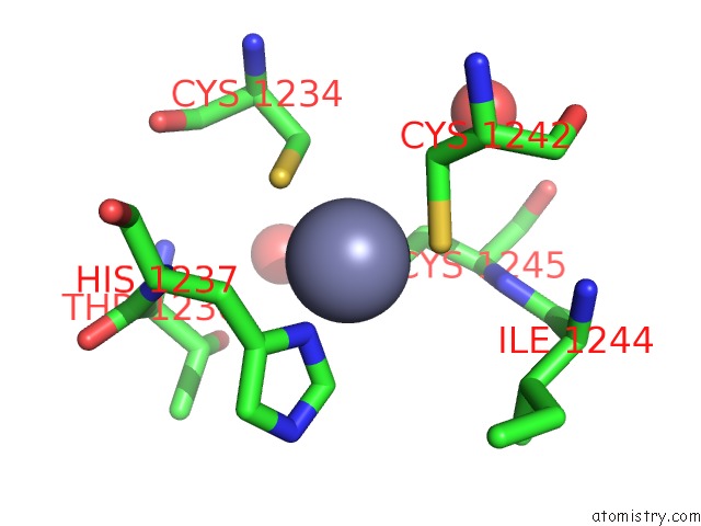
Mono view
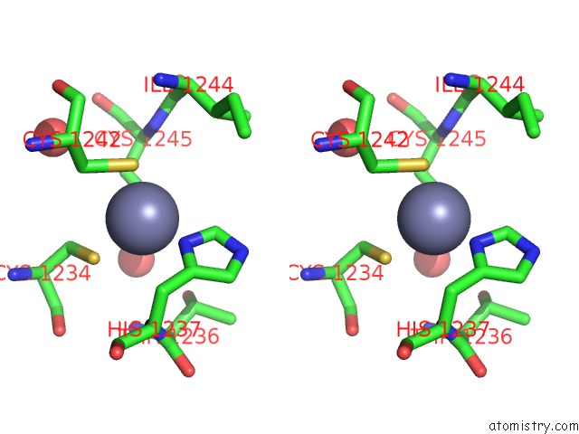
Stereo pair view
Mono view
Stereo pair view
A full contact list of Zinc with other atoms in the Zn binding
site number 1 of Crystal Structure of Tankyrase 1 with 355 within 5.0Å range:
|
Zinc binding site 2 out of 2 in 4tos
Go back to
Zinc binding site 2 out
of 2 in the Crystal Structure of Tankyrase 1 with 355
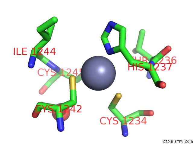
Mono view
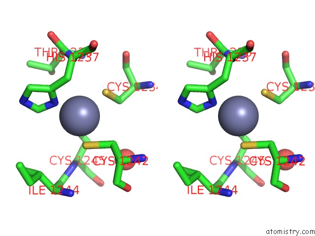
Stereo pair view
Mono view
Stereo pair view
A full contact list of Zinc with other atoms in the Zn binding
site number 2 of Crystal Structure of Tankyrase 1 with 355 within 5.0Å range:
|
Reference:
O.Kulak,
H.Chen,
B.Holohan,
X.Wu,
H.He,
D.Borek,
Z.Otwinowski,
K.Yamaguchi,
L.A.Garofalo,
Z.Ma,
W.Wright,
C.Chen,
J.W.Shay,
X.Zhang,
L.Lum.
Disruption of Wnt/ Beta-Catenin Signaling and Telomeric Shortening Are Inextricable Consequences of Tankyrase Inhibition in Human Cells. Mol.Cell.Biol. 2015.
ISSN: ESSN 1098-5549
PubMed: 25939383
DOI: 10.1128/MCB.00392-15
Page generated: Sun Oct 27 08:28:55 2024
ISSN: ESSN 1098-5549
PubMed: 25939383
DOI: 10.1128/MCB.00392-15
Last articles
Zn in 9MJ5Zn in 9HNW
Zn in 9G0L
Zn in 9FNE
Zn in 9DZN
Zn in 9E0I
Zn in 9D32
Zn in 9DAK
Zn in 8ZXC
Zn in 8ZUF