Zinc »
PDB 4rmg-4rvo »
4row »
Zinc in PDB 4row: The Crystal Structure of Novel APOBEC3G CD2 Head-to-Tail Dimer Suggests the Binding Mode of Full-Length APOBEC3G to Hiv-1 Ssdna
Protein crystallography data
The structure of The Crystal Structure of Novel APOBEC3G CD2 Head-to-Tail Dimer Suggests the Binding Mode of Full-Length APOBEC3G to Hiv-1 Ssdna, PDB code: 4row
was solved by
X.Lu,
T.Zhang,
Z.Xu,
S.Liu,
B.Zhao,
W.Lan,
C.Wang,
J.Ding,
C.Cao,
with X-Ray Crystallography technique. A brief refinement statistics is given in the table below:
| Resolution Low / High (Å) | 35.31 / 1.70 |
| Space group | P 2 21 21 |
| Cell size a, b, c (Å), α, β, γ (°) | 35.308, 70.007, 82.347, 90.00, 90.00, 90.00 |
| R / Rfree (%) | 14.7 / 18.8 |
Zinc Binding Sites:
The binding sites of Zinc atom in the The Crystal Structure of Novel APOBEC3G CD2 Head-to-Tail Dimer Suggests the Binding Mode of Full-Length APOBEC3G to Hiv-1 Ssdna
(pdb code 4row). This binding sites where shown within
5.0 Angstroms radius around Zinc atom.
In total only one binding site of Zinc was determined in the The Crystal Structure of Novel APOBEC3G CD2 Head-to-Tail Dimer Suggests the Binding Mode of Full-Length APOBEC3G to Hiv-1 Ssdna, PDB code: 4row:
In total only one binding site of Zinc was determined in the The Crystal Structure of Novel APOBEC3G CD2 Head-to-Tail Dimer Suggests the Binding Mode of Full-Length APOBEC3G to Hiv-1 Ssdna, PDB code: 4row:
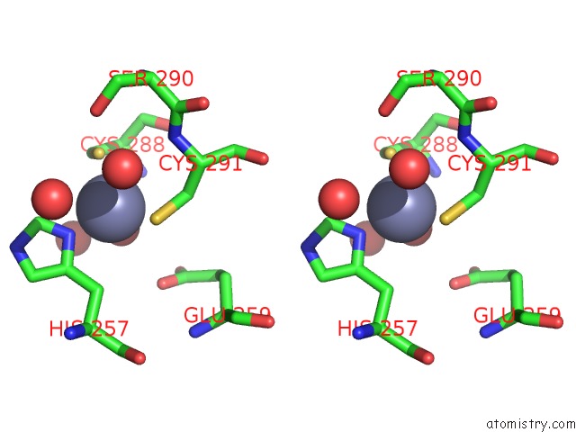
Zinc binding site 1 out of 1 in 4row
Go back to
Zinc binding site 1 out
of 1 in the The Crystal Structure of Novel APOBEC3G CD2 Head-to-Tail Dimer Suggests the Binding Mode of Full-Length APOBEC3G to Hiv-1 Ssdna
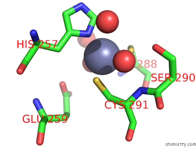
Mono view
Stereo pair view
Mono view
Stereo pair view
A full contact list of Zinc with other atoms in the Zn binding
site number 1 of The Crystal Structure of Novel APOBEC3G CD2 Head-to-Tail Dimer Suggests the Binding Mode of Full-Length APOBEC3G to Hiv-1 Ssdna within 5.0Å range:
|
Reference:
X.Lu,
T.Zhang,
Z.Xu,
S.Liu,
B.Zhao,
W.Lan,
C.Wang,
J.Ding,
C.Cao.
Crystal Structure of Dna Cytidine Deaminase ABOBEC3G Catalytic Domain Suggests A Binding Mode of Full-Length Enzyme to Single-Stranded Dna To Be Published.
Page generated: Wed Aug 20 22:13:22 2025
Last articles
Zn in 5IY5Zn in 5IX2
Zn in 5IX1
Zn in 5IX0
Zn in 5IWG
Zn in 5IWA
Zn in 5IT5
Zn in 5IW5
Zn in 5IQK
Zn in 5IT9