Zinc »
PDB 4qsb-4r6t »
4qy3 »
Zinc in PDB 4qy3: The Crystal Structure of the Complex of Hcaii with An Ortho- Substituted Benzoic Acid
Enzymatic activity of The Crystal Structure of the Complex of Hcaii with An Ortho- Substituted Benzoic Acid
All present enzymatic activity of The Crystal Structure of the Complex of Hcaii with An Ortho- Substituted Benzoic Acid:
4.2.1.1;
4.2.1.1;
Protein crystallography data
The structure of The Crystal Structure of the Complex of Hcaii with An Ortho- Substituted Benzoic Acid, PDB code: 4qy3
was solved by
K.D'ambrosio,
G.De Simone,
with X-Ray Crystallography technique. A brief refinement statistics is given in the table below:
| Resolution Low / High (Å) | 50.00 / 1.50 |
| Space group | P 1 21 1 |
| Cell size a, b, c (Å), α, β, γ (°) | 42.150, 41.530, 72.180, 90.00, 104.52, 90.00 |
| R / Rfree (%) | 16.3 / 18.4 |
Other elements in 4qy3:
The structure of The Crystal Structure of the Complex of Hcaii with An Ortho- Substituted Benzoic Acid also contains other interesting chemical elements:
| Mercury | (Hg) | 1 atom |
Zinc Binding Sites:
The binding sites of Zinc atom in the The Crystal Structure of the Complex of Hcaii with An Ortho- Substituted Benzoic Acid
(pdb code 4qy3). This binding sites where shown within
5.0 Angstroms radius around Zinc atom.
In total only one binding site of Zinc was determined in the The Crystal Structure of the Complex of Hcaii with An Ortho- Substituted Benzoic Acid, PDB code: 4qy3:
In total only one binding site of Zinc was determined in the The Crystal Structure of the Complex of Hcaii with An Ortho- Substituted Benzoic Acid, PDB code: 4qy3:
Zinc binding site 1 out of 1 in 4qy3
Go back to
Zinc binding site 1 out
of 1 in the The Crystal Structure of the Complex of Hcaii with An Ortho- Substituted Benzoic Acid
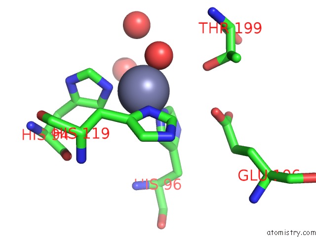
Mono view
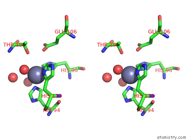
Stereo pair view
Mono view
Stereo pair view
A full contact list of Zinc with other atoms in the Zn binding
site number 1 of The Crystal Structure of the Complex of Hcaii with An Ortho- Substituted Benzoic Acid within 5.0Å range:
|
Reference:
K.D'ambrosio,
S.Carradori,
S.M.Monti,
M.Buonanno,
D.Secci,
D.Vullo,
C.T.Supuran,
G.De Simone.
Out of the Active Site Binding Pocket For Carbonic Anhydrase Inhibitors. Chem.Commun.(Camb.) 2014.
ISSN: ESSN 1364-548X
PubMed: 25407638
DOI: 10.1039/C4CC07320G
Page generated: Wed Aug 20 21:55:00 2025
ISSN: ESSN 1364-548X
PubMed: 25407638
DOI: 10.1039/C4CC07320G
Last articles
Zn in 5C97Zn in 5C7E
Zn in 5C8I
Zn in 5C84
Zn in 5C7F
Zn in 5C83
Zn in 5C66
Zn in 5C7D
Zn in 5C7C
Zn in 5C7B