Zinc »
PDB 4qia-4qqu »
4qpe »
Zinc in PDB 4qpe: Crystal Structure of Aminopeptidase N in Complex with N-Cyclohexyl-1, 2-Diaminoethylphosphonic Acid
Enzymatic activity of Crystal Structure of Aminopeptidase N in Complex with N-Cyclohexyl-1, 2-Diaminoethylphosphonic Acid
All present enzymatic activity of Crystal Structure of Aminopeptidase N in Complex with N-Cyclohexyl-1, 2-Diaminoethylphosphonic Acid:
3.4.11.2;
3.4.11.2;
Protein crystallography data
The structure of Crystal Structure of Aminopeptidase N in Complex with N-Cyclohexyl-1, 2-Diaminoethylphosphonic Acid, PDB code: 4qpe
was solved by
B.Nocek,
R.Mulligan,
L.Berlicki,
S.Vassilious,
A.Mucha,
A.Joachimiak,
with X-Ray Crystallography technique. A brief refinement statistics is given in the table below:
| Resolution Low / High (Å) | 28.06 / 2.00 |
| Space group | H 3 |
| Cell size a, b, c (Å), α, β, γ (°) | 224.495, 224.495, 57.951, 90.00, 90.00, 120.00 |
| R / Rfree (%) | 17.1 / 20.2 |
Zinc Binding Sites:
The binding sites of Zinc atom in the Crystal Structure of Aminopeptidase N in Complex with N-Cyclohexyl-1, 2-Diaminoethylphosphonic Acid
(pdb code 4qpe). This binding sites where shown within
5.0 Angstroms radius around Zinc atom.
In total only one binding site of Zinc was determined in the Crystal Structure of Aminopeptidase N in Complex with N-Cyclohexyl-1, 2-Diaminoethylphosphonic Acid, PDB code: 4qpe:
In total only one binding site of Zinc was determined in the Crystal Structure of Aminopeptidase N in Complex with N-Cyclohexyl-1, 2-Diaminoethylphosphonic Acid, PDB code: 4qpe:
Zinc binding site 1 out of 1 in 4qpe
Go back to
Zinc binding site 1 out
of 1 in the Crystal Structure of Aminopeptidase N in Complex with N-Cyclohexyl-1, 2-Diaminoethylphosphonic Acid
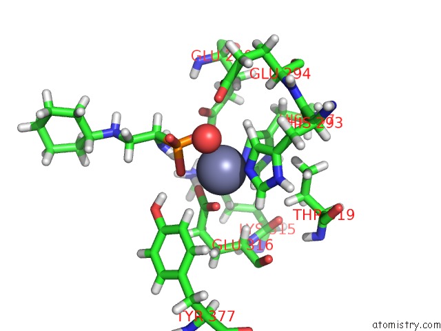
Mono view
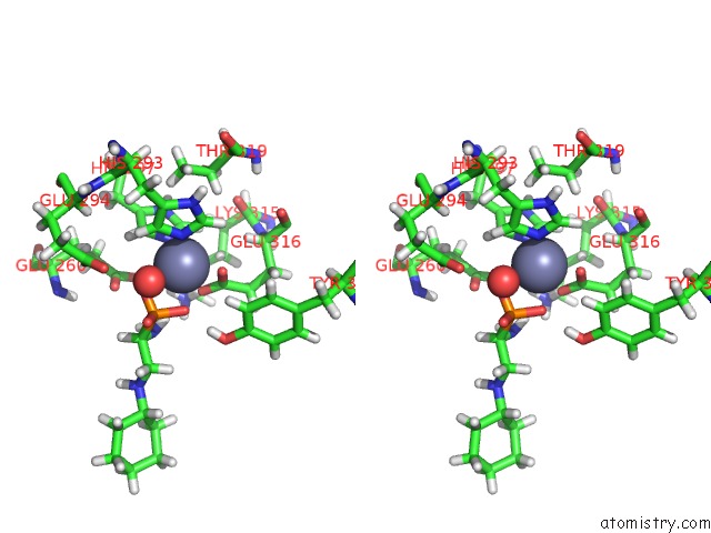
Stereo pair view
Mono view
Stereo pair view
A full contact list of Zinc with other atoms in the Zn binding
site number 1 of Crystal Structure of Aminopeptidase N in Complex with N-Cyclohexyl-1, 2-Diaminoethylphosphonic Acid within 5.0Å range:
|
Reference:
S.Vassiliou,
E.Weglarz-Tomczak,
L.Berlicki,
M.Paweczak,
B.Nocek,
R.Mulligan,
A.Joachimiak,
A.Mucha.
Structure-Guided, Single-Point Modifications in the Phosphinic Dipeptide Structure Yield Highly Potent and Selective Inhibitors of Neutral Aminopeptidases. J.Med.Chem. V. 57 8140 2014.
ISSN: ISSN 0022-2623
PubMed: 25192493
DOI: 10.1021/JM501071F
Page generated: Sun Oct 27 06:42:35 2024
ISSN: ISSN 0022-2623
PubMed: 25192493
DOI: 10.1021/JM501071F
Last articles
Zn in 9MJ5Zn in 9HNW
Zn in 9G0L
Zn in 9FNE
Zn in 9DZN
Zn in 9E0I
Zn in 9D32
Zn in 9DAK
Zn in 8ZXC
Zn in 8ZUF