Zinc »
PDB 4q47-4q8w »
4q8r »
Zinc in PDB 4q8r: Crystal Structure of A Phosphate Binding Protein (Pbp-1) From Clostridium Perfringens
Protein crystallography data
The structure of Crystal Structure of A Phosphate Binding Protein (Pbp-1) From Clostridium Perfringens, PDB code: 4q8r
was solved by
D.Gonzalez,
M.Richez,
C.Bergonzi,
E.Chabriere,
M.Elias,
with X-Ray Crystallography technique. A brief refinement statistics is given in the table below:
| Resolution Low / High (Å) | 37.56 / 1.65 |
| Space group | F 2 2 2 |
| Cell size a, b, c (Å), α, β, γ (°) | 70.780, 105.730, 146.450, 90.00, 90.00, 90.00 |
| R / Rfree (%) | 13.2 / 17.8 |
Zinc Binding Sites:
The binding sites of Zinc atom in the Crystal Structure of A Phosphate Binding Protein (Pbp-1) From Clostridium Perfringens
(pdb code 4q8r). This binding sites where shown within
5.0 Angstroms radius around Zinc atom.
In total 4 binding sites of Zinc where determined in the Crystal Structure of A Phosphate Binding Protein (Pbp-1) From Clostridium Perfringens, PDB code: 4q8r:
Jump to Zinc binding site number: 1; 2; 3; 4;
In total 4 binding sites of Zinc where determined in the Crystal Structure of A Phosphate Binding Protein (Pbp-1) From Clostridium Perfringens, PDB code: 4q8r:
Jump to Zinc binding site number: 1; 2; 3; 4;
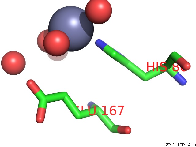
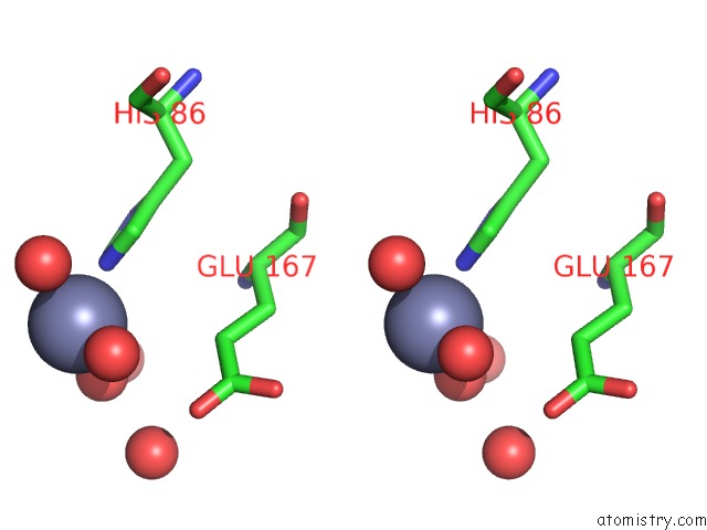
Zinc binding site 1 out of 4 in 4q8r
Go back to
Zinc binding site 1 out
of 4 in the Crystal Structure of A Phosphate Binding Protein (Pbp-1) From Clostridium Perfringens
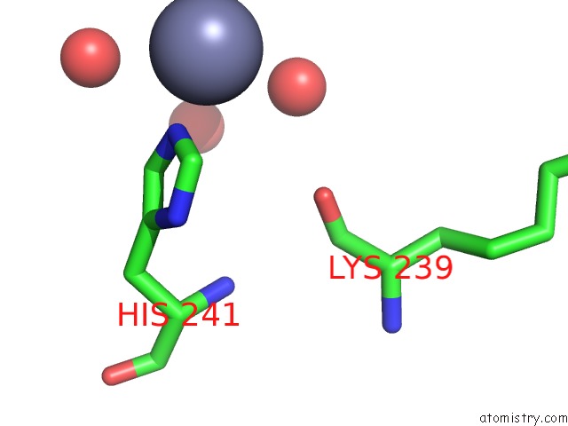
Mono view
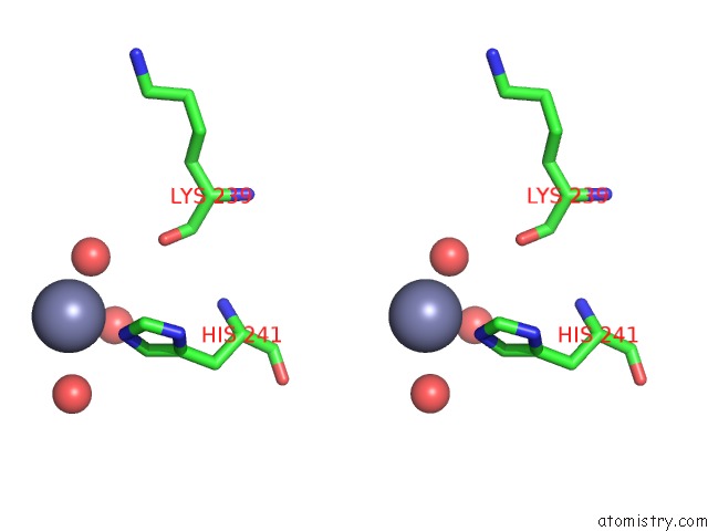
Stereo pair view
Mono view
Stereo pair view
A full contact list of Zinc with other atoms in the Zn binding
site number 1 of Crystal Structure of A Phosphate Binding Protein (Pbp-1) From Clostridium Perfringens within 5.0Å range:
|
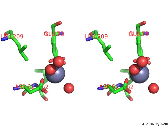
Zinc binding site 2 out of 4 in 4q8r
Go back to
Zinc binding site 2 out
of 4 in the Crystal Structure of A Phosphate Binding Protein (Pbp-1) From Clostridium Perfringens
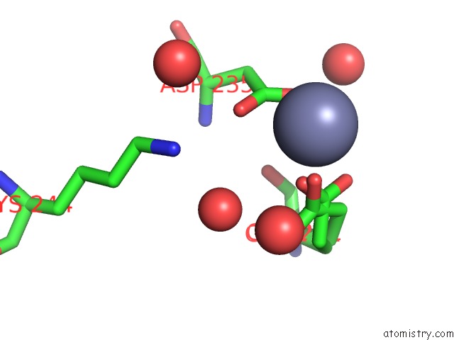
Mono view
Stereo pair view
Mono view
Stereo pair view
A full contact list of Zinc with other atoms in the Zn binding
site number 2 of Crystal Structure of A Phosphate Binding Protein (Pbp-1) From Clostridium Perfringens within 5.0Å range:
|
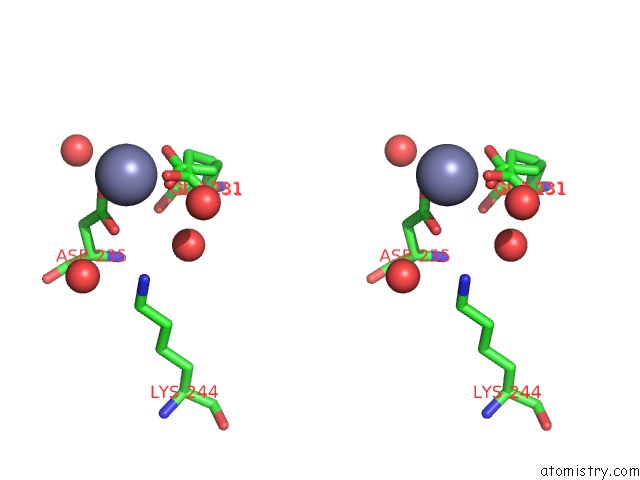
Zinc binding site 3 out of 4 in 4q8r
Go back to
Zinc binding site 3 out
of 4 in the Crystal Structure of A Phosphate Binding Protein (Pbp-1) From Clostridium Perfringens
Mono view
Stereo pair view
Mono view
Stereo pair view
A full contact list of Zinc with other atoms in the Zn binding
site number 3 of Crystal Structure of A Phosphate Binding Protein (Pbp-1) From Clostridium Perfringens within 5.0Å range:
|
Zinc binding site 4 out of 4 in 4q8r
Go back to
Zinc binding site 4 out
of 4 in the Crystal Structure of A Phosphate Binding Protein (Pbp-1) From Clostridium Perfringens

Mono view
Stereo pair view
Mono view
Stereo pair view
A full contact list of Zinc with other atoms in the Zn binding
site number 4 of Crystal Structure of A Phosphate Binding Protein (Pbp-1) From Clostridium Perfringens within 5.0Å range:
|
Reference:
D.Gonzalez,
M.Richez,
C.Bergonzi,
E.Chabriere,
M.Elias.
Crystal Structure of the Phosphate-Binding Protein (Pbp-1) of An Abc-Type Phosphate Transporter From Clostridium Perfringens. Sci Rep V. 4 6636 2014.
ISSN: ESSN 2045-2322
PubMed: 25338617
DOI: 10.1038/SREP06636
Page generated: Sun Oct 27 06:21:44 2024
ISSN: ESSN 2045-2322
PubMed: 25338617
DOI: 10.1038/SREP06636
Last articles
Zn in 9MJ5Zn in 9HNW
Zn in 9G0L
Zn in 9FNE
Zn in 9DZN
Zn in 9E0I
Zn in 9D32
Zn in 9DAK
Zn in 8ZXC
Zn in 8ZUF