Zinc »
PDB 4pa5-4ppe »
4pf9 »
Zinc in PDB 4pf9: Crystal Structure of Insulin Degrading Enzyme Complexed with Inhibitor
Enzymatic activity of Crystal Structure of Insulin Degrading Enzyme Complexed with Inhibitor
All present enzymatic activity of Crystal Structure of Insulin Degrading Enzyme Complexed with Inhibitor:
3.4.24.56;
3.4.24.56;
Protein crystallography data
The structure of Crystal Structure of Insulin Degrading Enzyme Complexed with Inhibitor, PDB code: 4pf9
was solved by
Y.Wang,
S.Guo,
with X-Ray Crystallography technique. A brief refinement statistics is given in the table below:
| Resolution Low / High (Å) | 19.99 / 2.50 |
| Space group | P 1 21 1 |
| Cell size a, b, c (Å), α, β, γ (°) | 79.254, 116.419, 124.470, 90.00, 97.60, 90.00 |
| R / Rfree (%) | 19.5 / 25.3 |
Zinc Binding Sites:
The binding sites of Zinc atom in the Crystal Structure of Insulin Degrading Enzyme Complexed with Inhibitor
(pdb code 4pf9). This binding sites where shown within
5.0 Angstroms radius around Zinc atom.
In total 2 binding sites of Zinc where determined in the Crystal Structure of Insulin Degrading Enzyme Complexed with Inhibitor, PDB code: 4pf9:
Jump to Zinc binding site number: 1; 2;
In total 2 binding sites of Zinc where determined in the Crystal Structure of Insulin Degrading Enzyme Complexed with Inhibitor, PDB code: 4pf9:
Jump to Zinc binding site number: 1; 2;
Zinc binding site 1 out of 2 in 4pf9
Go back to
Zinc binding site 1 out
of 2 in the Crystal Structure of Insulin Degrading Enzyme Complexed with Inhibitor
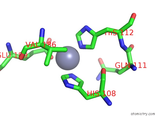
Mono view
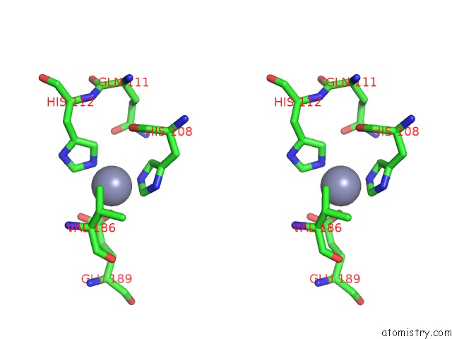
Stereo pair view
Mono view
Stereo pair view
A full contact list of Zinc with other atoms in the Zn binding
site number 1 of Crystal Structure of Insulin Degrading Enzyme Complexed with Inhibitor within 5.0Å range:
|
Zinc binding site 2 out of 2 in 4pf9
Go back to
Zinc binding site 2 out
of 2 in the Crystal Structure of Insulin Degrading Enzyme Complexed with Inhibitor
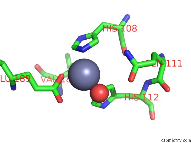
Mono view
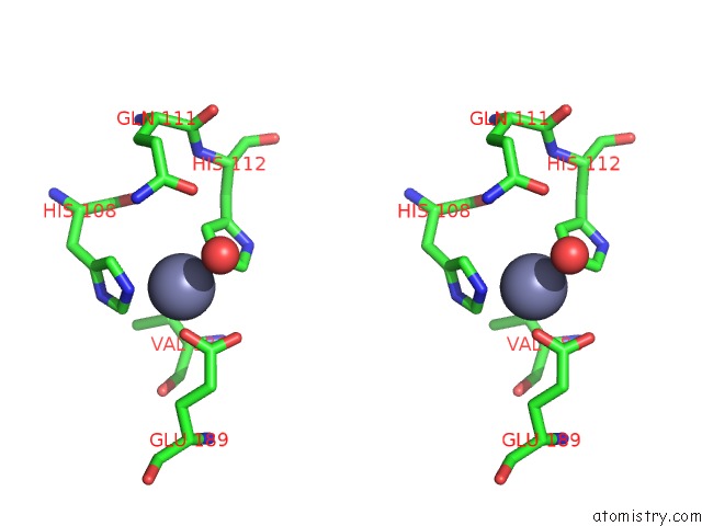
Stereo pair view
Mono view
Stereo pair view
A full contact list of Zinc with other atoms in the Zn binding
site number 2 of Crystal Structure of Insulin Degrading Enzyme Complexed with Inhibitor within 5.0Å range:
|
Reference:
T.B.Durham,
J.L.Toth,
V.J.Klimkowski,
J.X.Cao,
A.M.Siesky,
J.Alexander-Chacko,
G.Y.Wu,
J.T.Dixon,
J.E.Mcgee,
Y.Wang,
S.Y.Guo,
R.N.Cavitt,
J.Schindler,
S.J.Thibodeaux,
N.A.Calvert,
M.J.Coghlan,
D.K.Sindelar,
M.Christe,
V.V.Kiselyov,
M.D.Michael,
K.W.Sloop.
Dual Exosite-Binding Inhibitors of Insulin-Degrading Enzyme Challenge Its Role As the Primary Mediator of Insulin Clearance in Vivo. J.Biol.Chem. V. 290 20044 2015.
ISSN: ESSN 1083-351X
PubMed: 26085101
DOI: 10.1074/JBC.M115.638205
Page generated: Sun Oct 27 05:52:53 2024
ISSN: ESSN 1083-351X
PubMed: 26085101
DOI: 10.1074/JBC.M115.638205
Last articles
Zn in 9MJ5Zn in 9HNW
Zn in 9G0L
Zn in 9FNE
Zn in 9DZN
Zn in 9E0I
Zn in 9D32
Zn in 9DAK
Zn in 8ZXC
Zn in 8ZUF