Zinc »
PDB 4ox5-4p9e »
4p65 »
Zinc in PDB 4p65: Crystal Structure of An Cyclohexylalanine Substituted Insulin Analog.
Protein crystallography data
The structure of Crystal Structure of An Cyclohexylalanine Substituted Insulin Analog., PDB code: 4p65
was solved by
V.Pandyarajan,
Z.Wan,
M.A.Weiss,
with X-Ray Crystallography technique. A brief refinement statistics is given in the table below:
| Resolution Low / High (Å) | 42.30 / 1.50 |
| Space group | P 1 21 1 |
| Cell size a, b, c (Å), α, β, γ (°) | 46.040, 60.900, 59.300, 90.00, 112.25, 90.00 |
| R / Rfree (%) | 16 / 20.3 |
Other elements in 4p65:
The structure of Crystal Structure of An Cyclohexylalanine Substituted Insulin Analog. also contains other interesting chemical elements:
| Chlorine | (Cl) | 2 atoms |
Zinc Binding Sites:
The binding sites of Zinc atom in the Crystal Structure of An Cyclohexylalanine Substituted Insulin Analog.
(pdb code 4p65). This binding sites where shown within
5.0 Angstroms radius around Zinc atom.
In total 2 binding sites of Zinc where determined in the Crystal Structure of An Cyclohexylalanine Substituted Insulin Analog., PDB code: 4p65:
Jump to Zinc binding site number: 1; 2;
In total 2 binding sites of Zinc where determined in the Crystal Structure of An Cyclohexylalanine Substituted Insulin Analog., PDB code: 4p65:
Jump to Zinc binding site number: 1; 2;
Zinc binding site 1 out of 2 in 4p65
Go back to
Zinc binding site 1 out
of 2 in the Crystal Structure of An Cyclohexylalanine Substituted Insulin Analog.

Mono view
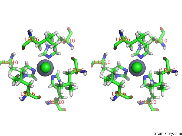
Stereo pair view
Mono view
Stereo pair view
A full contact list of Zinc with other atoms in the Zn binding
site number 1 of Crystal Structure of An Cyclohexylalanine Substituted Insulin Analog. within 5.0Å range:
|
Zinc binding site 2 out of 2 in 4p65
Go back to
Zinc binding site 2 out
of 2 in the Crystal Structure of An Cyclohexylalanine Substituted Insulin Analog.

Mono view
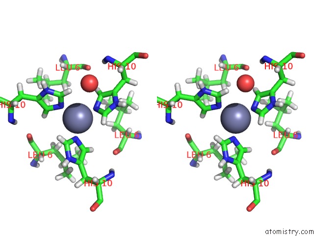
Stereo pair view
Mono view
Stereo pair view
A full contact list of Zinc with other atoms in the Zn binding
site number 2 of Crystal Structure of An Cyclohexylalanine Substituted Insulin Analog. within 5.0Å range:
|
Reference:
V.Pandyarajan,
B.J.Smith,
N.B.Phillips,
L.Whittaker,
G.P.Cox,
N.Wickramasinghe,
J.G.Menting,
Z.L.Wan,
J.Whittaker,
F.Ismail-Beigi,
M.C.Lawrence,
M.A.Weiss.
Aromatic Anchor at An Invariant Hormone-Receptor Interface. Function of Insulin Residue B24 with Application to Protein Design. J.Biol.Chem. 2014.
ISSN: ESSN 1083-351X
PubMed: 25305014
DOI: 10.1074/JBC.M114.608562
Page generated: Sun Oct 27 04:16:17 2024
ISSN: ESSN 1083-351X
PubMed: 25305014
DOI: 10.1074/JBC.M114.608562
Last articles
As in 3NLTAs in 3NRF
As in 3NLI
As in 3NLG
As in 3NLH
As in 3NLF
As in 3N6G
As in 3NLE
As in 3NLD
As in 3N6F