Zinc »
PDB 4o8x-4ojv »
4o8x »
Zinc in PDB 4o8x: Zinc-Bound RPN11 in Complex with RPN8
Protein crystallography data
The structure of Zinc-Bound RPN11 in Complex with RPN8, PDB code: 4o8x
was solved by
E.J.Worden,
C.Padovani,
A.Martin,
with X-Ray Crystallography technique. A brief refinement statistics is given in the table below:
| Resolution Low / High (Å) | 48.23 / 1.99 |
| Space group | P 43 21 2 |
| Cell size a, b, c (Å), α, β, γ (°) | 70.296, 70.296, 198.912, 90.00, 90.00, 90.00 |
| R / Rfree (%) | 16.1 / 19.5 |
Zinc Binding Sites:
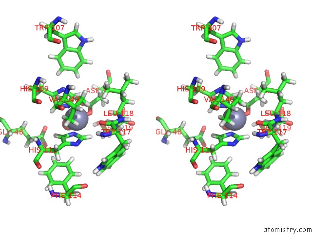
The binding sites of Zinc atom in the Zinc-Bound RPN11 in Complex with RPN8
(pdb code 4o8x). This binding sites where shown within
5.0 Angstroms radius around Zinc atom.
In total only one binding site of Zinc was determined in the Zinc-Bound RPN11 in Complex with RPN8, PDB code: 4o8x:
In total only one binding site of Zinc was determined in the Zinc-Bound RPN11 in Complex with RPN8, PDB code: 4o8x:
Zinc binding site 1 out of 1 in 4o8x
Go back to
Zinc binding site 1 out
of 1 in the Zinc-Bound RPN11 in Complex with RPN8

Mono view
Stereo pair view
Mono view
Stereo pair view
A full contact list of Zinc with other atoms in the Zn binding
site number 1 of Zinc-Bound RPN11 in Complex with RPN8 within 5.0Å range:
|
Reference:
E.J.Worden,
C.Padovani,
A.Martin.
Structure of the RPN11-RPN8 Dimer Reveals Mechanisms of Substrate Deubiquitination During Proteasomal Degradation. Nat.Struct.Mol.Biol. V. 21 220 2014.
ISSN: ISSN 1545-9993
PubMed: 24463465
DOI: 10.1038/NSMB.2771
Page generated: Sun Oct 27 03:37:04 2024
ISSN: ISSN 1545-9993
PubMed: 24463465
DOI: 10.1038/NSMB.2771
Last articles
Zn in 9MJ5Zn in 9HNW
Zn in 9G0L
Zn in 9FNE
Zn in 9DZN
Zn in 9E0I
Zn in 9D32
Zn in 9DAK
Zn in 8ZXC
Zn in 8ZUF