Zinc »
PDB 4n4e-4ngr »
4n9v »
Zinc in PDB 4n9v: High Resolution X-Ray Structure of Urate Oxidase in Complex with 8- Azaxanthine
Enzymatic activity of High Resolution X-Ray Structure of Urate Oxidase in Complex with 8- Azaxanthine
All present enzymatic activity of High Resolution X-Ray Structure of Urate Oxidase in Complex with 8- Azaxanthine:
1.7.3.3;
1.7.3.3;
Protein crystallography data
The structure of High Resolution X-Ray Structure of Urate Oxidase in Complex with 8- Azaxanthine, PDB code: 4n9v
was solved by
E.Oksanen,
M.P.Blakeley,
M.Budayova-Spano,
with X-Ray Crystallography technique. A brief refinement statistics is given in the table below:
| Resolution Low / High (Å) | 35.16 / 1.10 |
| Space group | I 2 2 2 |
| Cell size a, b, c (Å), α, β, γ (°) | 79.800, 95.080, 104.490, 90.00, 90.00, 90.00 |
| R / Rfree (%) | 13.9 / 15.3 |
Other elements in 4n9v:
The structure of High Resolution X-Ray Structure of Urate Oxidase in Complex with 8- Azaxanthine also contains other interesting chemical elements:
| Chlorine | (Cl) | 1 atom |
| Sodium | (Na) | 1 atom |
Zinc Binding Sites:
The binding sites of Zinc atom in the High Resolution X-Ray Structure of Urate Oxidase in Complex with 8- Azaxanthine
(pdb code 4n9v). This binding sites where shown within
5.0 Angstroms radius around Zinc atom.
In total 2 binding sites of Zinc where determined in the High Resolution X-Ray Structure of Urate Oxidase in Complex with 8- Azaxanthine, PDB code: 4n9v:
Jump to Zinc binding site number: 1; 2;
In total 2 binding sites of Zinc where determined in the High Resolution X-Ray Structure of Urate Oxidase in Complex with 8- Azaxanthine, PDB code: 4n9v:
Jump to Zinc binding site number: 1; 2;
Zinc binding site 1 out of 2 in 4n9v
Go back to
Zinc binding site 1 out
of 2 in the High Resolution X-Ray Structure of Urate Oxidase in Complex with 8- Azaxanthine
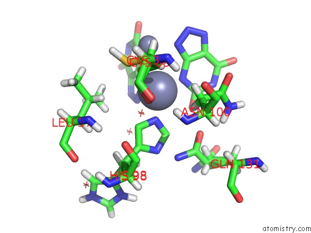
Mono view
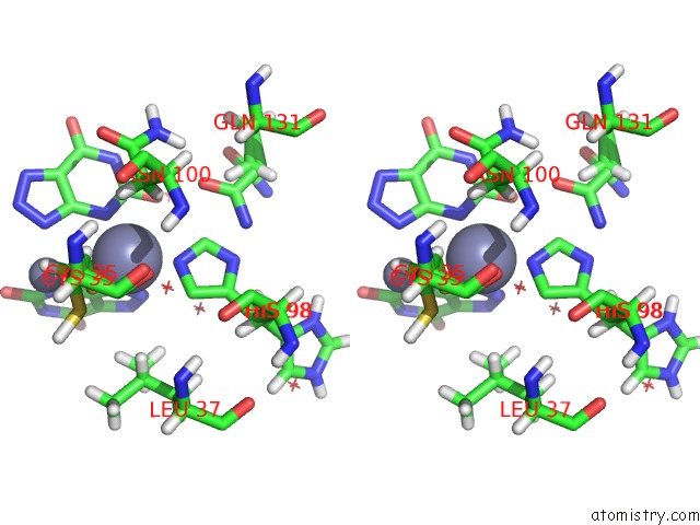
Stereo pair view
Mono view
Stereo pair view
A full contact list of Zinc with other atoms in the Zn binding
site number 1 of High Resolution X-Ray Structure of Urate Oxidase in Complex with 8- Azaxanthine within 5.0Å range:
|
Zinc binding site 2 out of 2 in 4n9v
Go back to
Zinc binding site 2 out
of 2 in the High Resolution X-Ray Structure of Urate Oxidase in Complex with 8- Azaxanthine
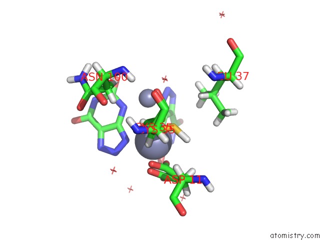
Mono view
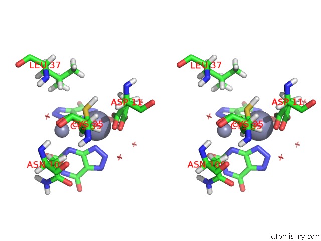
Stereo pair view
Mono view
Stereo pair view
A full contact list of Zinc with other atoms in the Zn binding
site number 2 of High Resolution X-Ray Structure of Urate Oxidase in Complex with 8- Azaxanthine within 5.0Å range:
|
Reference:
E.Oksanen,
M.P.Blakeley,
M.El-Hajji,
U.Ryde,
M.Budayova-Spano.
The Neutron Structure of Urate Oxidase Resolves A Long-Standing Mechanistic Conundrum and Reveals Unexpected Changes in Protonation. Plos One V. 9 86651 2014.
ISSN: ESSN 1932-6203
PubMed: 24466188
DOI: 10.1371/JOURNAL.PONE.0086651
Page generated: Sun Oct 27 03:02:39 2024
ISSN: ESSN 1932-6203
PubMed: 24466188
DOI: 10.1371/JOURNAL.PONE.0086651
Last articles
Zn in 9MJ5Zn in 9HNW
Zn in 9G0L
Zn in 9FNE
Zn in 9DZN
Zn in 9E0I
Zn in 9D32
Zn in 9DAK
Zn in 8ZXC
Zn in 8ZUF