Zinc »
PDB 4mti-4n3w »
4mut »
Zinc in PDB 4mut: Crystal Structure of Vancomycin Resistance D,D-Dipeptidase/D,D- Pentapeptidase Vanxyc D59S Mutant in Complex with D-Alanine
Protein crystallography data
The structure of Crystal Structure of Vancomycin Resistance D,D-Dipeptidase/D,D- Pentapeptidase Vanxyc D59S Mutant in Complex with D-Alanine, PDB code: 4mut
was solved by
P.J.Stogios,
E.Evdokimova,
D.Meziane-Cherif,
R.Di Leo,
V.Yim,
P.Courvalin,
A.Savchenko,
W.F.Anderson,
Center For Structural Genomics Ofinfectious Diseases (Csgid),
with X-Ray Crystallography technique. A brief refinement statistics is given in the table below:
| Resolution Low / High (Å) | 34.57 / 2.25 |
| Space group | P 1 |
| Cell size a, b, c (Å), α, β, γ (°) | 44.674, 45.214, 63.014, 86.69, 77.20, 63.88 |
| R / Rfree (%) | 16.1 / 18.5 |
Other elements in 4mut:
The structure of Crystal Structure of Vancomycin Resistance D,D-Dipeptidase/D,D- Pentapeptidase Vanxyc D59S Mutant in Complex with D-Alanine also contains other interesting chemical elements:
| Chlorine | (Cl) | 1 atom |
Zinc Binding Sites:
The binding sites of Zinc atom in the Crystal Structure of Vancomycin Resistance D,D-Dipeptidase/D,D- Pentapeptidase Vanxyc D59S Mutant in Complex with D-Alanine
(pdb code 4mut). This binding sites where shown within
5.0 Angstroms radius around Zinc atom.
In total 2 binding sites of Zinc where determined in the Crystal Structure of Vancomycin Resistance D,D-Dipeptidase/D,D- Pentapeptidase Vanxyc D59S Mutant in Complex with D-Alanine, PDB code: 4mut:
Jump to Zinc binding site number: 1; 2;
In total 2 binding sites of Zinc where determined in the Crystal Structure of Vancomycin Resistance D,D-Dipeptidase/D,D- Pentapeptidase Vanxyc D59S Mutant in Complex with D-Alanine, PDB code: 4mut:
Jump to Zinc binding site number: 1; 2;
Zinc binding site 1 out of 2 in 4mut
Go back to
Zinc binding site 1 out
of 2 in the Crystal Structure of Vancomycin Resistance D,D-Dipeptidase/D,D- Pentapeptidase Vanxyc D59S Mutant in Complex with D-Alanine
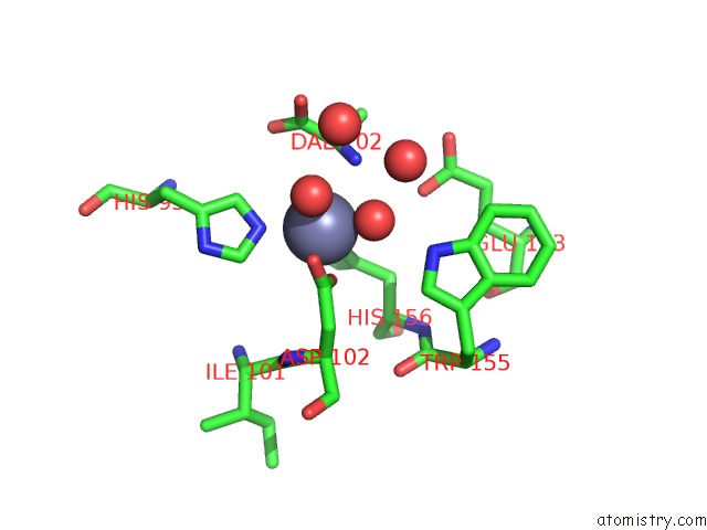
Mono view
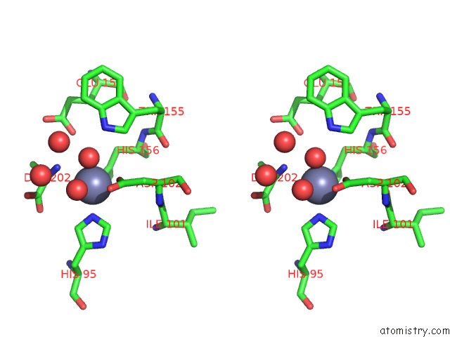
Stereo pair view
Mono view
Stereo pair view
A full contact list of Zinc with other atoms in the Zn binding
site number 1 of Crystal Structure of Vancomycin Resistance D,D-Dipeptidase/D,D- Pentapeptidase Vanxyc D59S Mutant in Complex with D-Alanine within 5.0Å range:
|
Zinc binding site 2 out of 2 in 4mut
Go back to
Zinc binding site 2 out
of 2 in the Crystal Structure of Vancomycin Resistance D,D-Dipeptidase/D,D- Pentapeptidase Vanxyc D59S Mutant in Complex with D-Alanine
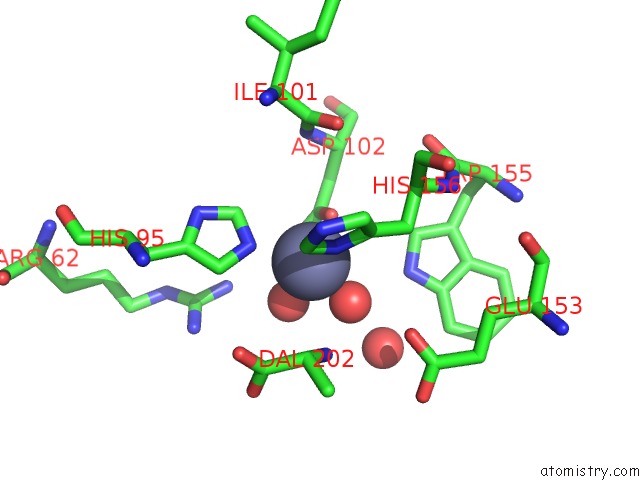
Mono view
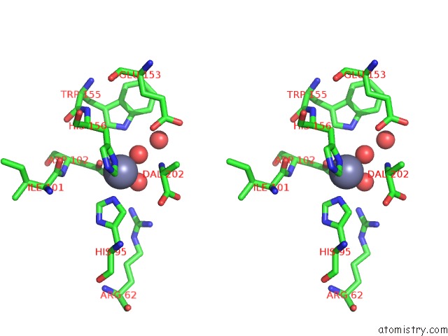
Stereo pair view
Mono view
Stereo pair view
A full contact list of Zinc with other atoms in the Zn binding
site number 2 of Crystal Structure of Vancomycin Resistance D,D-Dipeptidase/D,D- Pentapeptidase Vanxyc D59S Mutant in Complex with D-Alanine within 5.0Å range:
|
Reference:
D.Meziane-Cherif,
P.J.Stogios,
E.Evdokimova,
A.Savchenko,
P.Courvalin.
Structural Basis For the Evolution of Vancomycin Resistance D,D-Peptidases. Proc.Natl.Acad.Sci.Usa V. 111 5872 2014.
ISSN: ISSN 0027-8424
PubMed: 24711382
DOI: 10.1073/PNAS.1402259111
Page generated: Sun Oct 27 02:49:44 2024
ISSN: ISSN 0027-8424
PubMed: 24711382
DOI: 10.1073/PNAS.1402259111
Last articles
Zn in 9MJ5Zn in 9HNW
Zn in 9G0L
Zn in 9FNE
Zn in 9DZN
Zn in 9E0I
Zn in 9D32
Zn in 9DAK
Zn in 8ZXC
Zn in 8ZUF