Zinc »
PDB 4m3p-4mi0 »
4mg3 »
Zinc in PDB 4mg3: Crystal Structural Analysis of 2A Protease From Coxsackievirus A16
Enzymatic activity of Crystal Structural Analysis of 2A Protease From Coxsackievirus A16
All present enzymatic activity of Crystal Structural Analysis of 2A Protease From Coxsackievirus A16:
3.4.22.29;
3.4.22.29;
Protein crystallography data
The structure of Crystal Structural Analysis of 2A Protease From Coxsackievirus A16, PDB code: 4mg3
was solved by
Y.Sun,
X.Wang,
M.Dang,
S.Yuan,
with X-Ray Crystallography technique. A brief refinement statistics is given in the table below:
| Resolution Low / High (Å) | 35.47 / 1.80 |
| Space group | H 3 2 |
| Cell size a, b, c (Å), α, β, γ (°) | 101.271, 101.271, 193.914, 90.00, 90.00, 120.00 |
| R / Rfree (%) | 17.3 / 20.3 |
Zinc Binding Sites:
The binding sites of Zinc atom in the Crystal Structural Analysis of 2A Protease From Coxsackievirus A16
(pdb code 4mg3). This binding sites where shown within
5.0 Angstroms radius around Zinc atom.
In total 2 binding sites of Zinc where determined in the Crystal Structural Analysis of 2A Protease From Coxsackievirus A16, PDB code: 4mg3:
Jump to Zinc binding site number: 1; 2;
In total 2 binding sites of Zinc where determined in the Crystal Structural Analysis of 2A Protease From Coxsackievirus A16, PDB code: 4mg3:
Jump to Zinc binding site number: 1; 2;
Zinc binding site 1 out of 2 in 4mg3
Go back to
Zinc binding site 1 out
of 2 in the Crystal Structural Analysis of 2A Protease From Coxsackievirus A16
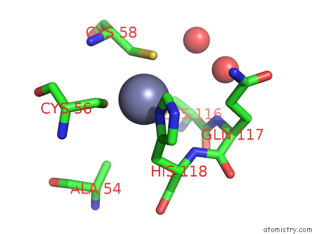
Mono view
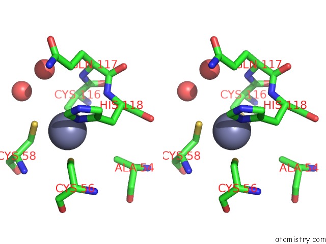
Stereo pair view
Mono view
Stereo pair view
A full contact list of Zinc with other atoms in the Zn binding
site number 1 of Crystal Structural Analysis of 2A Protease From Coxsackievirus A16 within 5.0Å range:
|
Zinc binding site 2 out of 2 in 4mg3
Go back to
Zinc binding site 2 out
of 2 in the Crystal Structural Analysis of 2A Protease From Coxsackievirus A16
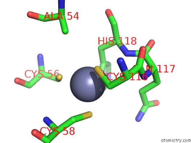
Mono view
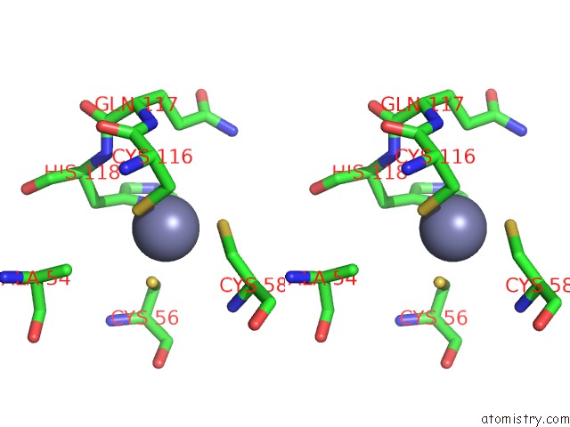
Stereo pair view
Mono view
Stereo pair view
A full contact list of Zinc with other atoms in the Zn binding
site number 2 of Crystal Structural Analysis of 2A Protease From Coxsackievirus A16 within 5.0Å range:
|
Reference:
Y.Sun,
X.Wang,
S.Yuan,
M.Dang,
X.Li,
X.C.Zhang,
Z.Rao.
An Open Conformation Determined By A Structural Switch For 2A Protease From Coxsackievirus A16. Protein Cell V. 4 782 2013.
ISSN: ISSN 1674-800X
PubMed: 24026848
DOI: 10.1007/S13238-013-3914-Z
Page generated: Sun Oct 27 02:27:08 2024
ISSN: ISSN 1674-800X
PubMed: 24026848
DOI: 10.1007/S13238-013-3914-Z
Last articles
Zn in 9MJ5Zn in 9HNW
Zn in 9G0L
Zn in 9FNE
Zn in 9DZN
Zn in 9E0I
Zn in 9D32
Zn in 9DAK
Zn in 8ZXC
Zn in 8ZUF