Zinc »
PDB 4lnb-4m3o »
4lqg »
Zinc in PDB 4lqg: X-Ray Structure of Human Glutamate Carboxypeptidase II (Gcpii) in Complex with A Phosphoramidate Inhibitor CTT1056
Enzymatic activity of X-Ray Structure of Human Glutamate Carboxypeptidase II (Gcpii) in Complex with A Phosphoramidate Inhibitor CTT1056
All present enzymatic activity of X-Ray Structure of Human Glutamate Carboxypeptidase II (Gcpii) in Complex with A Phosphoramidate Inhibitor CTT1056:
3.4.17.21;
3.4.17.21;
Protein crystallography data
The structure of X-Ray Structure of Human Glutamate Carboxypeptidase II (Gcpii) in Complex with A Phosphoramidate Inhibitor CTT1056, PDB code: 4lqg
was solved by
C.Barinka,
L.Skultetyova,
with X-Ray Crystallography technique. A brief refinement statistics is given in the table below:
| Resolution Low / High (Å) | 19.51 / 1.77 |
| Space group | I 2 2 2 |
| Cell size a, b, c (Å), α, β, γ (°) | 101.900, 130.233, 158.252, 90.00, 90.00, 90.00 |
| R / Rfree (%) | 15.7 / 18.2 |
Other elements in 4lqg:
The structure of X-Ray Structure of Human Glutamate Carboxypeptidase II (Gcpii) in Complex with A Phosphoramidate Inhibitor CTT1056 also contains other interesting chemical elements:
| Fluorine | (F) | 1 atom |
| Calcium | (Ca) | 1 atom |
| Chlorine | (Cl) | 1 atom |
Zinc Binding Sites:
The binding sites of Zinc atom in the X-Ray Structure of Human Glutamate Carboxypeptidase II (Gcpii) in Complex with A Phosphoramidate Inhibitor CTT1056
(pdb code 4lqg). This binding sites where shown within
5.0 Angstroms radius around Zinc atom.
In total 2 binding sites of Zinc where determined in the X-Ray Structure of Human Glutamate Carboxypeptidase II (Gcpii) in Complex with A Phosphoramidate Inhibitor CTT1056, PDB code: 4lqg:
Jump to Zinc binding site number: 1; 2;
In total 2 binding sites of Zinc where determined in the X-Ray Structure of Human Glutamate Carboxypeptidase II (Gcpii) in Complex with A Phosphoramidate Inhibitor CTT1056, PDB code: 4lqg:
Jump to Zinc binding site number: 1; 2;
Zinc binding site 1 out of 2 in 4lqg
Go back to
Zinc binding site 1 out
of 2 in the X-Ray Structure of Human Glutamate Carboxypeptidase II (Gcpii) in Complex with A Phosphoramidate Inhibitor CTT1056
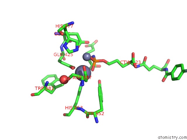
Mono view
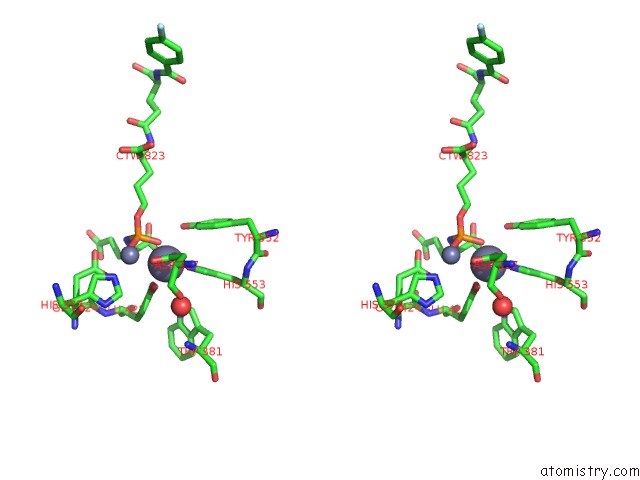
Stereo pair view
Mono view
Stereo pair view
A full contact list of Zinc with other atoms in the Zn binding
site number 1 of X-Ray Structure of Human Glutamate Carboxypeptidase II (Gcpii) in Complex with A Phosphoramidate Inhibitor CTT1056 within 5.0Å range:
|
Zinc binding site 2 out of 2 in 4lqg
Go back to
Zinc binding site 2 out
of 2 in the X-Ray Structure of Human Glutamate Carboxypeptidase II (Gcpii) in Complex with A Phosphoramidate Inhibitor CTT1056
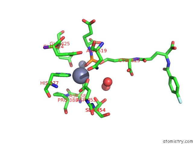
Mono view
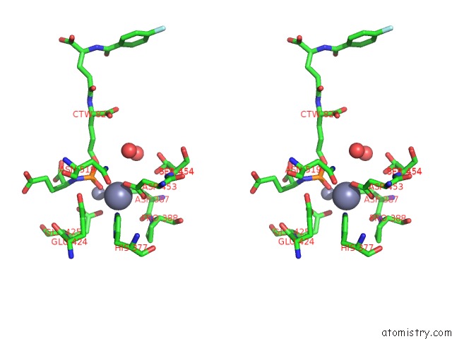
Stereo pair view
Mono view
Stereo pair view
A full contact list of Zinc with other atoms in the Zn binding
site number 2 of X-Ray Structure of Human Glutamate Carboxypeptidase II (Gcpii) in Complex with A Phosphoramidate Inhibitor CTT1056 within 5.0Å range:
|
Reference:
S.Dannoon,
T.Ganguly,
H.Cahaya,
J.G.Geruntho,
M.R.Hopkins,
M.Regan,
J.E.Blecha,
S.Jivan,
C.Barinka,
E.F.Jones,
H.F.Vanbrocklin,
C.E.Berkman.
Structure-Activity Relationship of 18F-Labeled Phosphoramidate Peptidomimetic Psma-Targeted Inhibitor Analogues For Pet Imaging of Prostate Cancer To Be Published.
Page generated: Sun Oct 27 02:04:41 2024
Last articles
Zn in 9MJ5Zn in 9HNW
Zn in 9G0L
Zn in 9FNE
Zn in 9DZN
Zn in 9E0I
Zn in 9D32
Zn in 9DAK
Zn in 8ZXC
Zn in 8ZUF