Zinc »
PDB 4l61-4leq »
4l64 »
Zinc in PDB 4l64: Crystal Structure of the Candida Albicans Methionine Synthase in Complex with 5-Methyl-Tetrahydrofolate
Enzymatic activity of Crystal Structure of the Candida Albicans Methionine Synthase in Complex with 5-Methyl-Tetrahydrofolate
All present enzymatic activity of Crystal Structure of the Candida Albicans Methionine Synthase in Complex with 5-Methyl-Tetrahydrofolate:
2.1.1.14;
2.1.1.14;
Protein crystallography data
The structure of Crystal Structure of the Candida Albicans Methionine Synthase in Complex with 5-Methyl-Tetrahydrofolate, PDB code: 4l64
was solved by
D.Ubhi,
J.D.Robertus,
with X-Ray Crystallography technique. A brief refinement statistics is given in the table below:
| Resolution Low / High (Å) | 49.61 / 2.18 |
| Space group | P 21 21 21 |
| Cell size a, b, c (Å), α, β, γ (°) | 76.862, 99.124, 100.719, 90.00, 90.00, 90.00 |
| R / Rfree (%) | 18.1 / 24.3 |
Zinc Binding Sites:
The binding sites of Zinc atom in the Crystal Structure of the Candida Albicans Methionine Synthase in Complex with 5-Methyl-Tetrahydrofolate
(pdb code 4l64). This binding sites where shown within
5.0 Angstroms radius around Zinc atom.
In total only one binding site of Zinc was determined in the Crystal Structure of the Candida Albicans Methionine Synthase in Complex with 5-Methyl-Tetrahydrofolate, PDB code: 4l64:
In total only one binding site of Zinc was determined in the Crystal Structure of the Candida Albicans Methionine Synthase in Complex with 5-Methyl-Tetrahydrofolate, PDB code: 4l64:
Zinc binding site 1 out of 1 in 4l64
Go back to
Zinc binding site 1 out
of 1 in the Crystal Structure of the Candida Albicans Methionine Synthase in Complex with 5-Methyl-Tetrahydrofolate
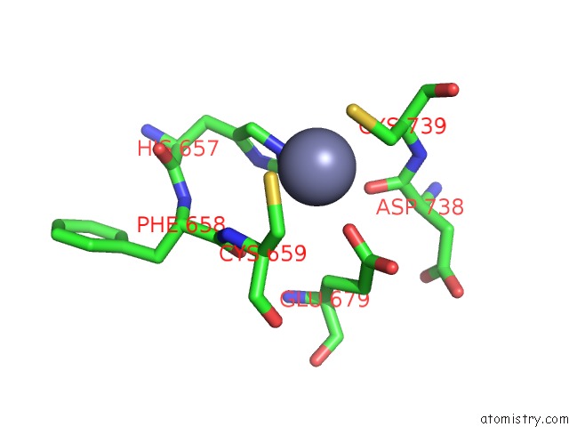
Mono view
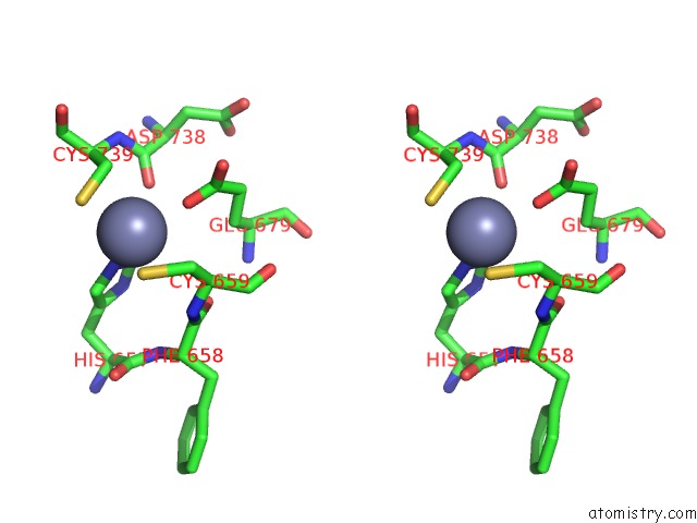
Stereo pair view
Mono view
Stereo pair view
A full contact list of Zinc with other atoms in the Zn binding
site number 1 of Crystal Structure of the Candida Albicans Methionine Synthase in Complex with 5-Methyl-Tetrahydrofolate within 5.0Å range:
|
Reference:
D.Ubhi,
G.Kago,
A.F.Monzingo,
J.D.Robertus.
Structural Analysis of A Fungal Methionine Synthase with Substrates and Inhibitors. J.Mol.Biol. V. 426 1839 2014.
ISSN: ISSN 0022-2836
PubMed: 24524835
DOI: 10.1016/J.JMB.2014.02.006
Page generated: Sun Oct 27 01:36:14 2024
ISSN: ISSN 0022-2836
PubMed: 24524835
DOI: 10.1016/J.JMB.2014.02.006
Last articles
Zn in 9MJ5Zn in 9HNW
Zn in 9G0L
Zn in 9FNE
Zn in 9DZN
Zn in 9E0I
Zn in 9D32
Zn in 9DAK
Zn in 8ZXC
Zn in 8ZUF