Zinc »
PDB 4kyh-4l5z »
4l3k »
Zinc in PDB 4l3k: Crystal Structure of Sporosarcina Pasteurii Uree Bound to NI2+ and ZN2+
Protein crystallography data
The structure of Crystal Structure of Sporosarcina Pasteurii Uree Bound to NI2+ and ZN2+, PDB code: 4l3k
was solved by
B.Zambelli,
K.Banaszak,
A.Merloni,
A.Kiliszek,
W.R.Rypniewski,
S.Ciurli,
with X-Ray Crystallography technique. A brief refinement statistics is given in the table below:
| Resolution Low / High (Å) | 45.66 / 1.88 |
| Space group | P 63 |
| Cell size a, b, c (Å), α, β, γ (°) | 91.329, 91.329, 82.792, 90.00, 90.00, 120.00 |
| R / Rfree (%) | 17.1 / 20.7 |
Other elements in 4l3k:
The structure of Crystal Structure of Sporosarcina Pasteurii Uree Bound to NI2+ and ZN2+ also contains other interesting chemical elements:
| Nickel | (Ni) | 1 atom |
Zinc Binding Sites:
The binding sites of Zinc atom in the Crystal Structure of Sporosarcina Pasteurii Uree Bound to NI2+ and ZN2+
(pdb code 4l3k). This binding sites where shown within
5.0 Angstroms radius around Zinc atom.
In total only one binding site of Zinc was determined in the Crystal Structure of Sporosarcina Pasteurii Uree Bound to NI2+ and ZN2+, PDB code: 4l3k:
In total only one binding site of Zinc was determined in the Crystal Structure of Sporosarcina Pasteurii Uree Bound to NI2+ and ZN2+, PDB code: 4l3k:
Zinc binding site 1 out of 1 in 4l3k
Go back to
Zinc binding site 1 out
of 1 in the Crystal Structure of Sporosarcina Pasteurii Uree Bound to NI2+ and ZN2+
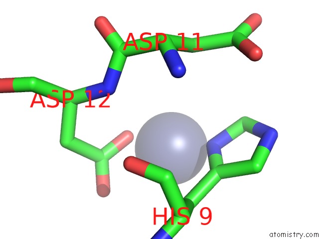
Mono view
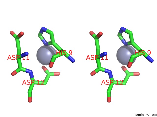
Stereo pair view
Mono view
Stereo pair view
A full contact list of Zinc with other atoms in the Zn binding
site number 1 of Crystal Structure of Sporosarcina Pasteurii Uree Bound to NI2+ and ZN2+ within 5.0Å range:
|
Reference:
B.Zambelli,
K.Banaszak,
A.Merloni,
A.Kiliszek,
W.Rypniewski,
S.Ciurli.
Selectivity of Ni(II) and Zn(II) Binding to Sporosarcina Pasteurii Uree, A Metallochaperone in the Urease Assembly: A Calorimetric and Crystallographic Study. J.Biol.Inorg.Chem. V. 18 1005 2013.
ISSN: ISSN 0949-8257
PubMed: 24126709
DOI: 10.1007/S00775-013-1049-6
Page generated: Sun Oct 27 01:33:10 2024
ISSN: ISSN 0949-8257
PubMed: 24126709
DOI: 10.1007/S00775-013-1049-6
Last articles
Zn in 9MJ5Zn in 9HNW
Zn in 9G0L
Zn in 9FNE
Zn in 9DZN
Zn in 9E0I
Zn in 9D32
Zn in 9DAK
Zn in 8ZXC
Zn in 8ZUF