Zinc »
PDB 4k4t-4k9a »
4k6m »
Zinc in PDB 4k6m: Crystal Structure of the Full-Length Japanese Encephalitis Virus NS5
Protein crystallography data
The structure of Crystal Structure of the Full-Length Japanese Encephalitis Virus NS5, PDB code: 4k6m
was solved by
G.Lu,
P.Gong,
with X-Ray Crystallography technique. A brief refinement statistics is given in the table below:
| Resolution Low / High (Å) | 43.22 / 2.60 |
| Space group | H 3 |
| Cell size a, b, c (Å), α, β, γ (°) | 272.293, 272.293, 177.248, 90.00, 90.00, 120.00 |
| R / Rfree (%) | 19.6 / 22.8 |
Zinc Binding Sites:
The binding sites of Zinc atom in the Crystal Structure of the Full-Length Japanese Encephalitis Virus NS5
(pdb code 4k6m). This binding sites where shown within
5.0 Angstroms radius around Zinc atom.
In total 4 binding sites of Zinc where determined in the Crystal Structure of the Full-Length Japanese Encephalitis Virus NS5, PDB code: 4k6m:
Jump to Zinc binding site number: 1; 2; 3; 4;
In total 4 binding sites of Zinc where determined in the Crystal Structure of the Full-Length Japanese Encephalitis Virus NS5, PDB code: 4k6m:
Jump to Zinc binding site number: 1; 2; 3; 4;
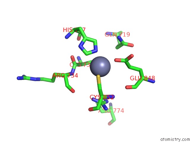
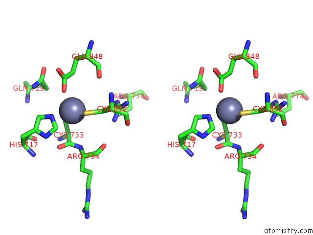
Zinc binding site 1 out of 4 in 4k6m
Go back to
Zinc binding site 1 out
of 4 in the Crystal Structure of the Full-Length Japanese Encephalitis Virus NS5
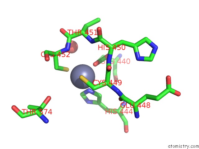
Mono view
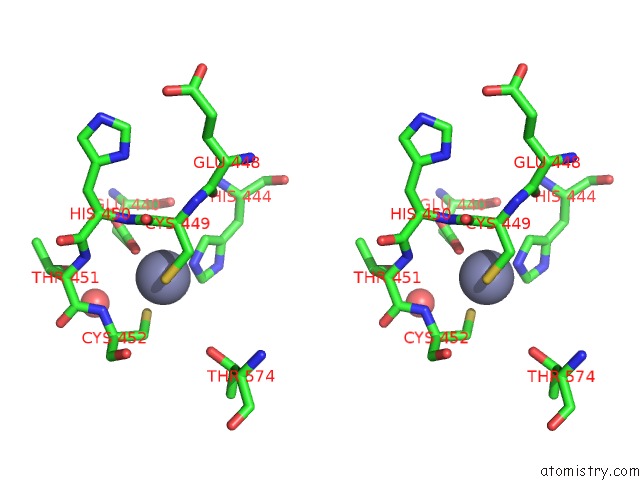
Stereo pair view
Mono view
Stereo pair view
A full contact list of Zinc with other atoms in the Zn binding
site number 1 of Crystal Structure of the Full-Length Japanese Encephalitis Virus NS5 within 5.0Å range:
|
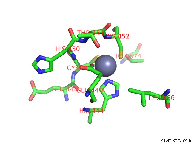
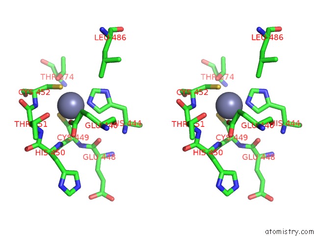
Zinc binding site 2 out of 4 in 4k6m
Go back to
Zinc binding site 2 out
of 4 in the Crystal Structure of the Full-Length Japanese Encephalitis Virus NS5
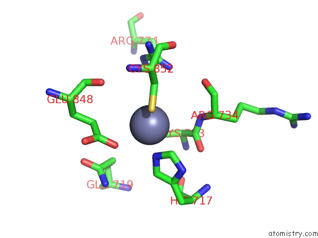
Mono view
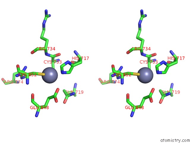
Stereo pair view
Mono view
Stereo pair view
A full contact list of Zinc with other atoms in the Zn binding
site number 2 of Crystal Structure of the Full-Length Japanese Encephalitis Virus NS5 within 5.0Å range:
|
Zinc binding site 3 out of 4 in 4k6m
Go back to
Zinc binding site 3 out
of 4 in the Crystal Structure of the Full-Length Japanese Encephalitis Virus NS5
Mono view
Stereo pair view
Mono view
Stereo pair view
A full contact list of Zinc with other atoms in the Zn binding
site number 3 of Crystal Structure of the Full-Length Japanese Encephalitis Virus NS5 within 5.0Å range:
|
Zinc binding site 4 out of 4 in 4k6m
Go back to
Zinc binding site 4 out
of 4 in the Crystal Structure of the Full-Length Japanese Encephalitis Virus NS5
Mono view
Stereo pair view
Mono view
Stereo pair view
A full contact list of Zinc with other atoms in the Zn binding
site number 4 of Crystal Structure of the Full-Length Japanese Encephalitis Virus NS5 within 5.0Å range:
|
Reference:
G.Lu,
P.Gong.
Crystal Structure of the Full-Length Japanese Encephalitis Virus NS5 Reveals A Conserved Methyltransferase-Polymerase Interface Plos Pathog. V. 9 03549 2013.
ISSN: ISSN 1553-7366
PubMed: 23950717
DOI: 10.1371/JOURNAL.PPAT.1003549
Page generated: Sun Oct 27 01:45:03 2024
ISSN: ISSN 1553-7366
PubMed: 23950717
DOI: 10.1371/JOURNAL.PPAT.1003549
Last articles
Zn in 9MJ5Zn in 9HNW
Zn in 9G0L
Zn in 9FNE
Zn in 9DZN
Zn in 9E0I
Zn in 9D32
Zn in 9DAK
Zn in 8ZXC
Zn in 8ZUF