Zinc »
PDB 4jsg-4k4s »
4jx4 »
Zinc in PDB 4jx4: Structure of the Carboxyl Transferase Domain From Rhizobium Etli Pyruvate Carboxylase
Enzymatic activity of Structure of the Carboxyl Transferase Domain From Rhizobium Etli Pyruvate Carboxylase
All present enzymatic activity of Structure of the Carboxyl Transferase Domain From Rhizobium Etli Pyruvate Carboxylase:
6.4.1.1;
6.4.1.1;
Protein crystallography data
The structure of Structure of the Carboxyl Transferase Domain From Rhizobium Etli Pyruvate Carboxylase, PDB code: 4jx4
was solved by
A.D.Lietzan,
M.St Maurice,
with X-Ray Crystallography technique. A brief refinement statistics is given in the table below:
| Resolution Low / High (Å) | 50.00 / 2.98 |
| Space group | P 21 21 21 |
| Cell size a, b, c (Å), α, β, γ (°) | 86.211, 157.227, 245.144, 90.00, 90.00, 90.00 |
| R / Rfree (%) | 21.4 / 24.8 |
Other elements in 4jx4:
The structure of Structure of the Carboxyl Transferase Domain From Rhizobium Etli Pyruvate Carboxylase also contains other interesting chemical elements:
| Chlorine | (Cl) | 2 atoms |
Zinc Binding Sites:
The binding sites of Zinc atom in the Structure of the Carboxyl Transferase Domain From Rhizobium Etli Pyruvate Carboxylase
(pdb code 4jx4). This binding sites where shown within
5.0 Angstroms radius around Zinc atom.
In total 4 binding sites of Zinc where determined in the Structure of the Carboxyl Transferase Domain From Rhizobium Etli Pyruvate Carboxylase, PDB code: 4jx4:
Jump to Zinc binding site number: 1; 2; 3; 4;
In total 4 binding sites of Zinc where determined in the Structure of the Carboxyl Transferase Domain From Rhizobium Etli Pyruvate Carboxylase, PDB code: 4jx4:
Jump to Zinc binding site number: 1; 2; 3; 4;
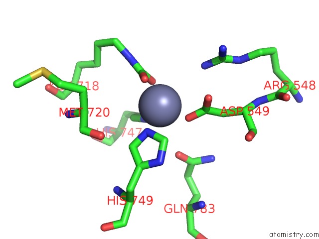
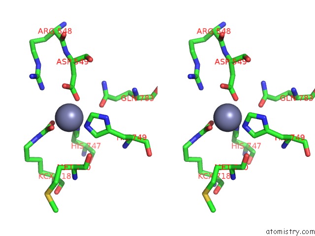
Zinc binding site 1 out of 4 in 4jx4
Go back to
Zinc binding site 1 out
of 4 in the Structure of the Carboxyl Transferase Domain From Rhizobium Etli Pyruvate Carboxylase
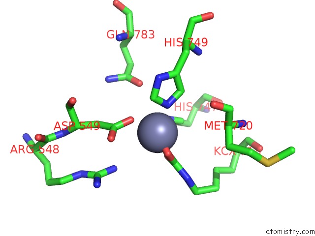
Mono view
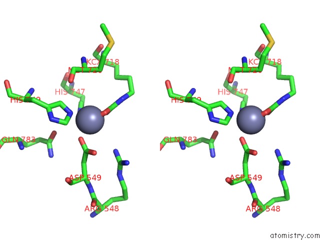
Stereo pair view
Mono view
Stereo pair view
A full contact list of Zinc with other atoms in the Zn binding
site number 1 of Structure of the Carboxyl Transferase Domain From Rhizobium Etli Pyruvate Carboxylase within 5.0Å range:
|
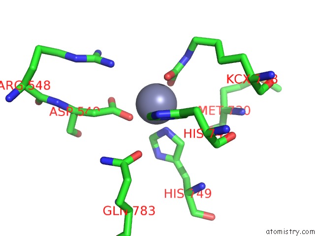
Zinc binding site 2 out of 4 in 4jx4
Go back to
Zinc binding site 2 out
of 4 in the Structure of the Carboxyl Transferase Domain From Rhizobium Etli Pyruvate Carboxylase
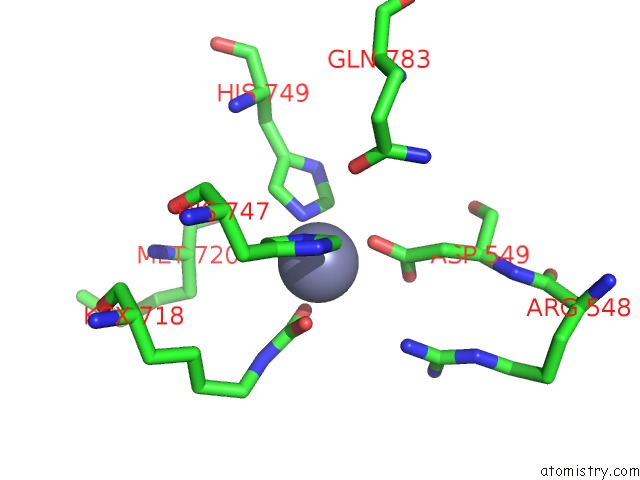
Mono view
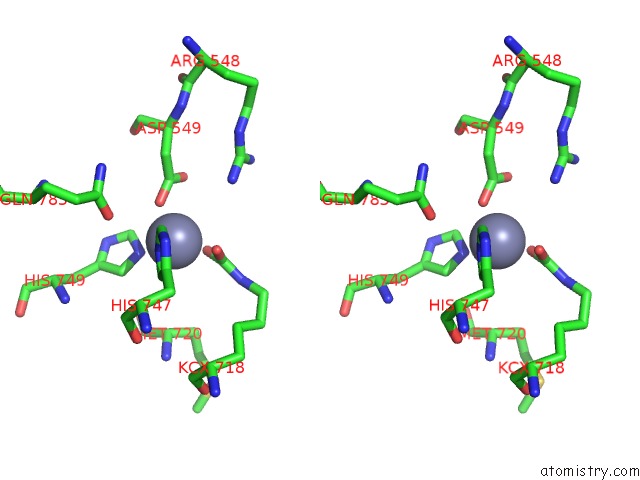
Stereo pair view
Mono view
Stereo pair view
A full contact list of Zinc with other atoms in the Zn binding
site number 2 of Structure of the Carboxyl Transferase Domain From Rhizobium Etli Pyruvate Carboxylase within 5.0Å range:
|
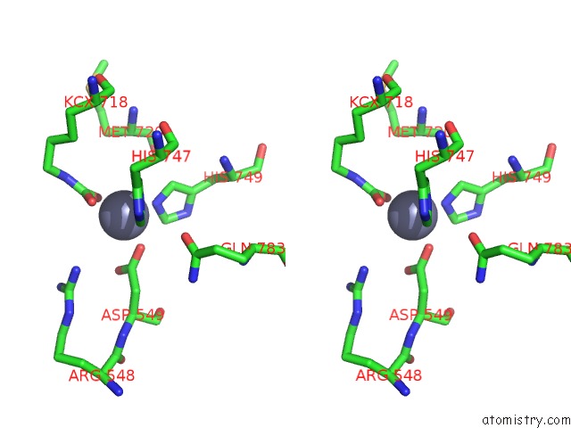
Zinc binding site 3 out of 4 in 4jx4
Go back to
Zinc binding site 3 out
of 4 in the Structure of the Carboxyl Transferase Domain From Rhizobium Etli Pyruvate Carboxylase
Mono view
Stereo pair view
Mono view
Stereo pair view
A full contact list of Zinc with other atoms in the Zn binding
site number 3 of Structure of the Carboxyl Transferase Domain From Rhizobium Etli Pyruvate Carboxylase within 5.0Å range:
|
Zinc binding site 4 out of 4 in 4jx4
Go back to
Zinc binding site 4 out
of 4 in the Structure of the Carboxyl Transferase Domain From Rhizobium Etli Pyruvate Carboxylase
Mono view
Stereo pair view
Mono view
Stereo pair view
A full contact list of Zinc with other atoms in the Zn binding
site number 4 of Structure of the Carboxyl Transferase Domain From Rhizobium Etli Pyruvate Carboxylase within 5.0Å range:
|
Reference:
A.D.Lietzan,
M.St Maurice.
A Substrate-Induced Biotin Binding Pocket in the Carboxyltransferase Domain of Pyruvate Carboxylase. J.Biol.Chem. V. 288 19915 2013.
ISSN: ISSN 0021-9258
PubMed: 23698000
DOI: 10.1074/JBC.M113.477828
Page generated: Sun Oct 27 01:30:43 2024
ISSN: ISSN 0021-9258
PubMed: 23698000
DOI: 10.1074/JBC.M113.477828
Last articles
Zn in 9MJ5Zn in 9HNW
Zn in 9G0L
Zn in 9FNE
Zn in 9DZN
Zn in 9E0I
Zn in 9D32
Zn in 9DAK
Zn in 8ZXC
Zn in 8ZUF