Zinc »
PDB 4ijd-4iwz »
4is9 »
Zinc in PDB 4is9: Crystal Structure of the Escherichia Coli Lpxc/L-161,240 Complex
Protein crystallography data
The structure of Crystal Structure of the Escherichia Coli Lpxc/L-161,240 Complex, PDB code: 4is9
was solved by
C.-J.Lee,
P.Zhou,
with X-Ray Crystallography technique. A brief refinement statistics is given in the table below:
| Resolution Low / High (Å) | 35.92 / 2.13 |
| Space group | H 3 2 |
| Cell size a, b, c (Å), α, β, γ (°) | 117.440, 117.440, 253.661, 90.00, 90.00, 120.00 |
| R / Rfree (%) | 21.6 / 24.7 |
Other elements in 4is9:
The structure of Crystal Structure of the Escherichia Coli Lpxc/L-161,240 Complex also contains other interesting chemical elements:
| Sodium | (Na) | 1 atom |
Zinc Binding Sites:
The binding sites of Zinc atom in the Crystal Structure of the Escherichia Coli Lpxc/L-161,240 Complex
(pdb code 4is9). This binding sites where shown within
5.0 Angstroms radius around Zinc atom.
In total 2 binding sites of Zinc where determined in the Crystal Structure of the Escherichia Coli Lpxc/L-161,240 Complex, PDB code: 4is9:
Jump to Zinc binding site number: 1; 2;
In total 2 binding sites of Zinc where determined in the Crystal Structure of the Escherichia Coli Lpxc/L-161,240 Complex, PDB code: 4is9:
Jump to Zinc binding site number: 1; 2;
Zinc binding site 1 out of 2 in 4is9
Go back to
Zinc binding site 1 out
of 2 in the Crystal Structure of the Escherichia Coli Lpxc/L-161,240 Complex
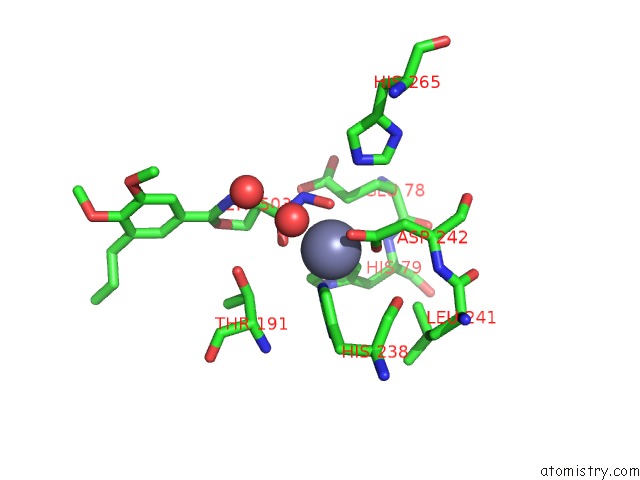
Mono view
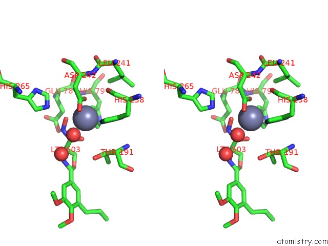
Stereo pair view
Mono view
Stereo pair view
A full contact list of Zinc with other atoms in the Zn binding
site number 1 of Crystal Structure of the Escherichia Coli Lpxc/L-161,240 Complex within 5.0Å range:
|
Zinc binding site 2 out of 2 in 4is9
Go back to
Zinc binding site 2 out
of 2 in the Crystal Structure of the Escherichia Coli Lpxc/L-161,240 Complex
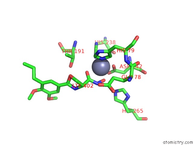
Mono view
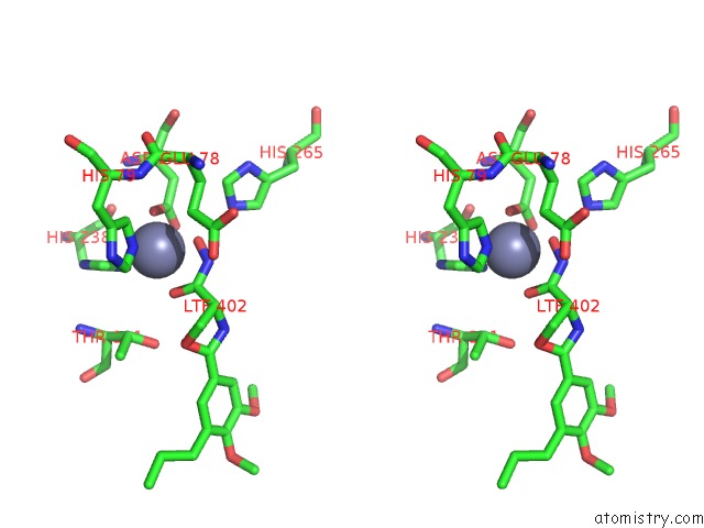
Stereo pair view
Mono view
Stereo pair view
A full contact list of Zinc with other atoms in the Zn binding
site number 2 of Crystal Structure of the Escherichia Coli Lpxc/L-161,240 Complex within 5.0Å range:
|
Reference:
C.J.Lee,
X.Liang,
R.Gopalaswamy,
J.Najeeb,
E.D.Ark,
E.J.Toone,
P.Zhou.
Structural Basis of the Promiscuous Inhibitor Susceptibility of Escherichia Coli Lpxc. Acs Chem.Biol. V. 9 237 2014.
ISSN: ISSN 1554-8929
PubMed: 24117400
DOI: 10.1021/CB400067G
Page generated: Sun Oct 27 00:51:35 2024
ISSN: ISSN 1554-8929
PubMed: 24117400
DOI: 10.1021/CB400067G
Last articles
Zn in 9MJ5Zn in 9HNW
Zn in 9G0L
Zn in 9FNE
Zn in 9DZN
Zn in 9E0I
Zn in 9D32
Zn in 9DAK
Zn in 8ZXC
Zn in 8ZUF