Zinc »
PDB 4ijd-4iwz »
4iou »
Zinc in PDB 4iou: Crystal Structure of the Hiv-1 Vif Binding, Catalytically Active Domain of APOBEC3F
Protein crystallography data
The structure of Crystal Structure of the Hiv-1 Vif Binding, Catalytically Active Domain of APOBEC3F, PDB code: 4iou
was solved by
M.Bohn,
S.M.D.Shandilya,
C.A.Schiffer,
with X-Ray Crystallography technique. A brief refinement statistics is given in the table below:
| Resolution Low / High (Å) | 36.46 / 2.75 |
| Space group | P 1 |
| Cell size a, b, c (Å), α, β, γ (°) | 50.790, 66.910, 75.640, 110.75, 94.41, 108.83 |
| R / Rfree (%) | 19.3 / 23.3 |
Zinc Binding Sites:
The binding sites of Zinc atom in the Crystal Structure of the Hiv-1 Vif Binding, Catalytically Active Domain of APOBEC3F
(pdb code 4iou). This binding sites where shown within
5.0 Angstroms radius around Zinc atom.
In total 4 binding sites of Zinc where determined in the Crystal Structure of the Hiv-1 Vif Binding, Catalytically Active Domain of APOBEC3F, PDB code: 4iou:
Jump to Zinc binding site number: 1; 2; 3; 4;
In total 4 binding sites of Zinc where determined in the Crystal Structure of the Hiv-1 Vif Binding, Catalytically Active Domain of APOBEC3F, PDB code: 4iou:
Jump to Zinc binding site number: 1; 2; 3; 4;
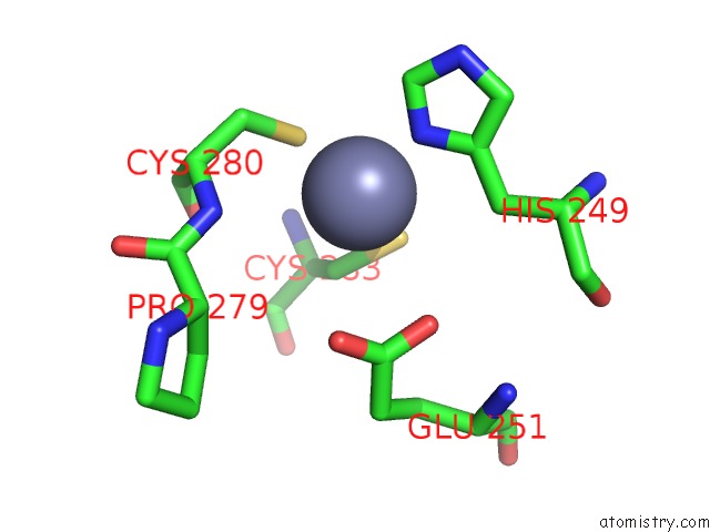
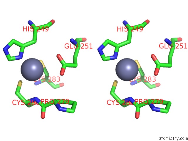
Zinc binding site 1 out of 4 in 4iou
Go back to
Zinc binding site 1 out
of 4 in the Crystal Structure of the Hiv-1 Vif Binding, Catalytically Active Domain of APOBEC3F
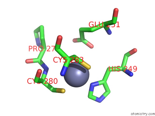
Mono view
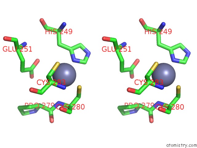
Stereo pair view
Mono view
Stereo pair view
A full contact list of Zinc with other atoms in the Zn binding
site number 1 of Crystal Structure of the Hiv-1 Vif Binding, Catalytically Active Domain of APOBEC3F within 5.0Å range:
|
Zinc binding site 2 out of 4 in 4iou
Go back to
Zinc binding site 2 out
of 4 in the Crystal Structure of the Hiv-1 Vif Binding, Catalytically Active Domain of APOBEC3F
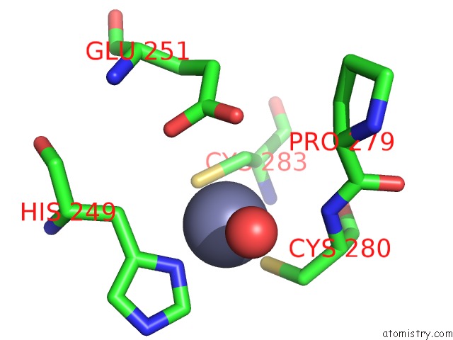
Mono view
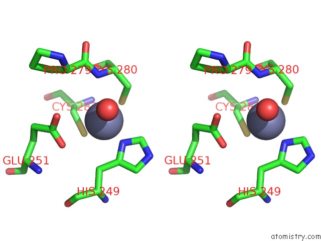
Stereo pair view
Mono view
Stereo pair view
A full contact list of Zinc with other atoms in the Zn binding
site number 2 of Crystal Structure of the Hiv-1 Vif Binding, Catalytically Active Domain of APOBEC3F within 5.0Å range:
|
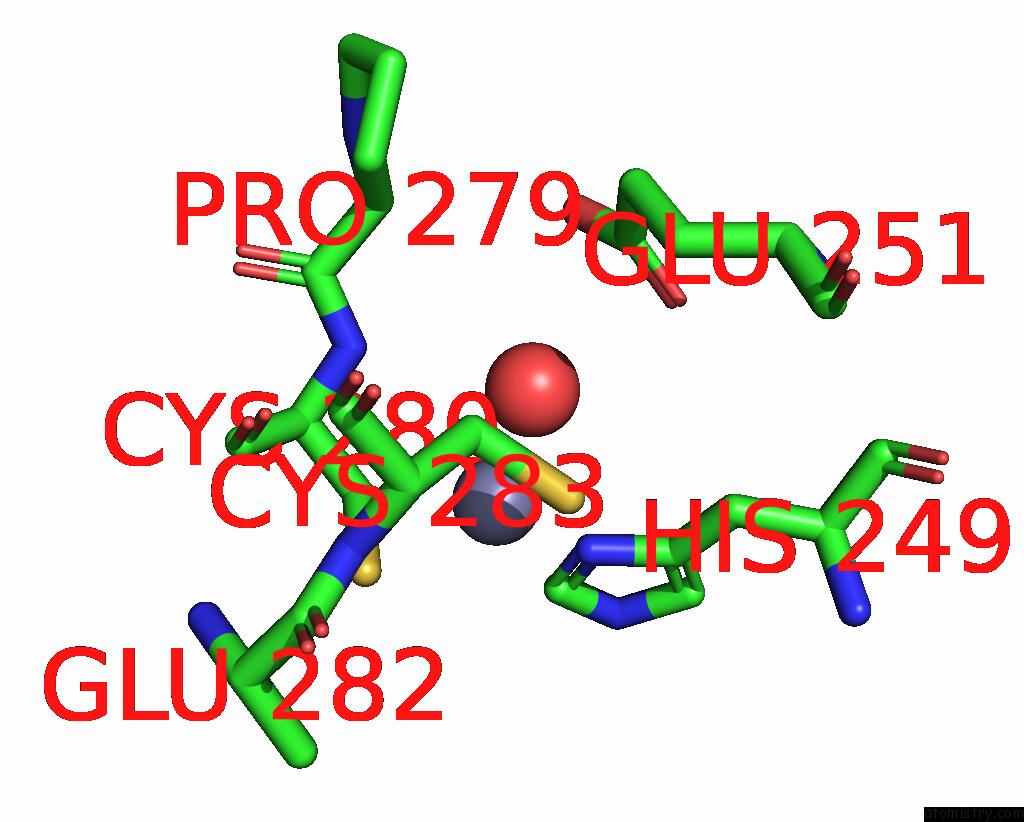
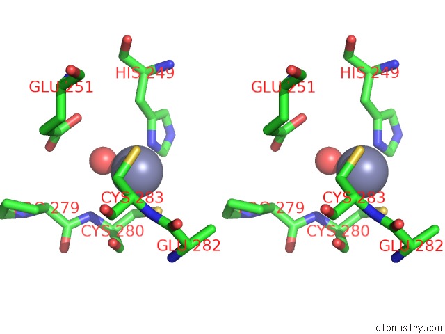
Zinc binding site 3 out of 4 in 4iou
Go back to
Zinc binding site 3 out
of 4 in the Crystal Structure of the Hiv-1 Vif Binding, Catalytically Active Domain of APOBEC3F
Mono view
Stereo pair view
Mono view
Stereo pair view
A full contact list of Zinc with other atoms in the Zn binding
site number 3 of Crystal Structure of the Hiv-1 Vif Binding, Catalytically Active Domain of APOBEC3F within 5.0Å range:
|
Zinc binding site 4 out of 4 in 4iou
Go back to
Zinc binding site 4 out
of 4 in the Crystal Structure of the Hiv-1 Vif Binding, Catalytically Active Domain of APOBEC3F
Mono view
Stereo pair view
Mono view
Stereo pair view
A full contact list of Zinc with other atoms in the Zn binding
site number 4 of Crystal Structure of the Hiv-1 Vif Binding, Catalytically Active Domain of APOBEC3F within 5.0Å range:
|
Reference:
M.F.Bohn,
S.M.Shandilya,
J.S.Albin,
T.Kouno,
B.D.Anderson,
R.M.Mcdougle,
M.A.Carpenter,
A.Rathore,
L.Evans,
A.N.Davis,
J.Zhang,
Y.Lu,
M.Somasundaran,
H.Matsuo,
R.S.Harris,
C.A.Schiffer.
Crystal Structure of the Dna Cytosine Deaminase APOBEC3F: the Catalytically Active and Hiv-1 Vif-Binding Domain. Structure V. 21 1042 2013.
ISSN: ISSN 0969-2126
PubMed: 23685212
DOI: 10.1016/J.STR.2013.04.010
Page generated: Sun Oct 27 00:50:05 2024
ISSN: ISSN 0969-2126
PubMed: 23685212
DOI: 10.1016/J.STR.2013.04.010
Last articles
Zn in 9MJ5Zn in 9HNW
Zn in 9G0L
Zn in 9FNE
Zn in 9DZN
Zn in 9E0I
Zn in 9D32
Zn in 9DAK
Zn in 8ZXC
Zn in 8ZUF