Zinc »
PDB 4hx2-4i9i »
4i9i »
Zinc in PDB 4i9i: Crystal Structure of Tankyrase 1 with Compound 4
Enzymatic activity of Crystal Structure of Tankyrase 1 with Compound 4
All present enzymatic activity of Crystal Structure of Tankyrase 1 with Compound 4:
2.4.2.30;
2.4.2.30;
Protein crystallography data
The structure of Crystal Structure of Tankyrase 1 with Compound 4, PDB code: 4i9i
was solved by
X.Huang,
with X-Ray Crystallography technique. A brief refinement statistics is given in the table below:
| Resolution Low / High (Å) | 50.00 / 2.40 |
| Space group | P 21 21 2 |
| Cell size a, b, c (Å), α, β, γ (°) | 158.622, 77.122, 84.507, 90.00, 90.00, 90.00 |
| R / Rfree (%) | 25 / 28.6 |
Zinc Binding Sites:
The binding sites of Zinc atom in the Crystal Structure of Tankyrase 1 with Compound 4
(pdb code 4i9i). This binding sites where shown within
5.0 Angstroms radius around Zinc atom.
In total 4 binding sites of Zinc where determined in the Crystal Structure of Tankyrase 1 with Compound 4, PDB code: 4i9i:
Jump to Zinc binding site number: 1; 2; 3; 4;
In total 4 binding sites of Zinc where determined in the Crystal Structure of Tankyrase 1 with Compound 4, PDB code: 4i9i:
Jump to Zinc binding site number: 1; 2; 3; 4;
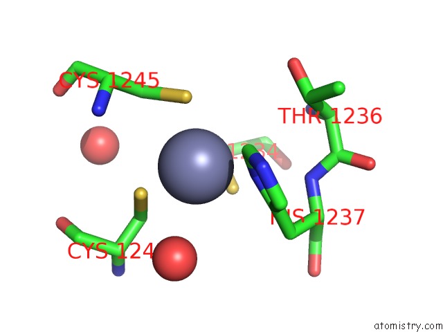
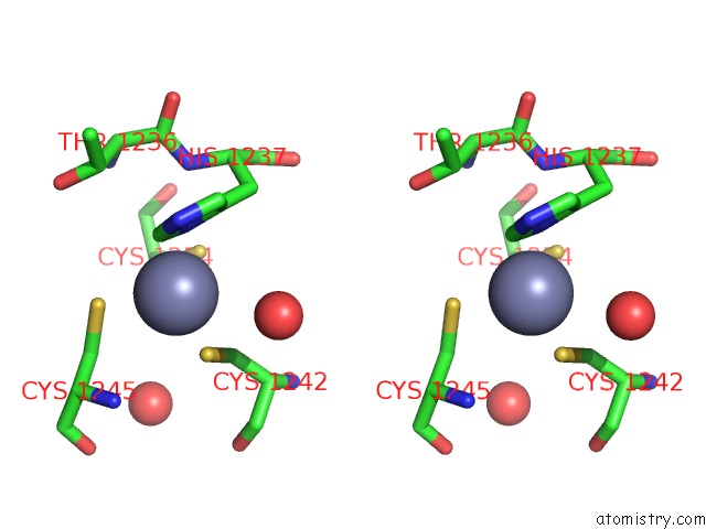
Zinc binding site 1 out of 4 in 4i9i
Go back to
Zinc binding site 1 out
of 4 in the Crystal Structure of Tankyrase 1 with Compound 4
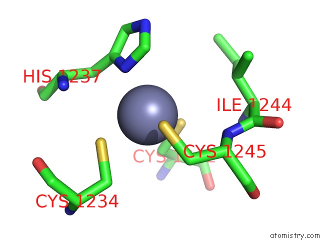
Mono view
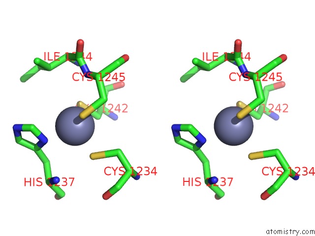
Stereo pair view
Mono view
Stereo pair view
A full contact list of Zinc with other atoms in the Zn binding
site number 1 of Crystal Structure of Tankyrase 1 with Compound 4 within 5.0Å range:
|
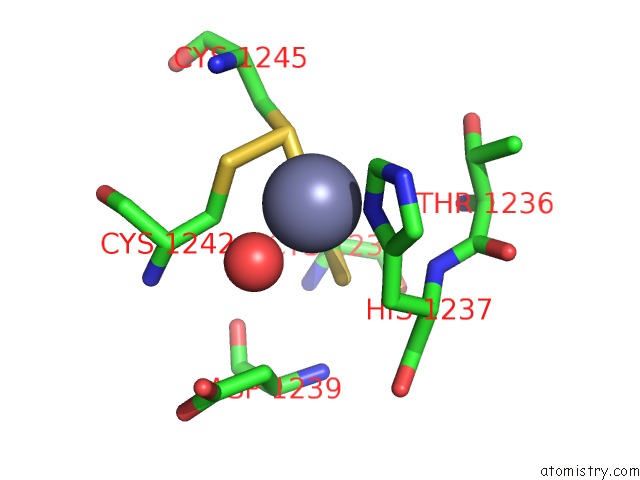
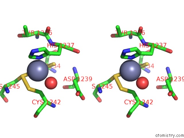
Zinc binding site 2 out of 4 in 4i9i
Go back to
Zinc binding site 2 out
of 4 in the Crystal Structure of Tankyrase 1 with Compound 4
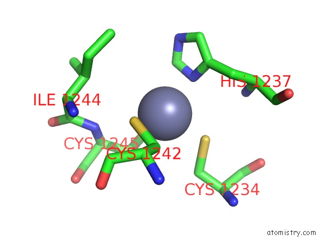
Mono view
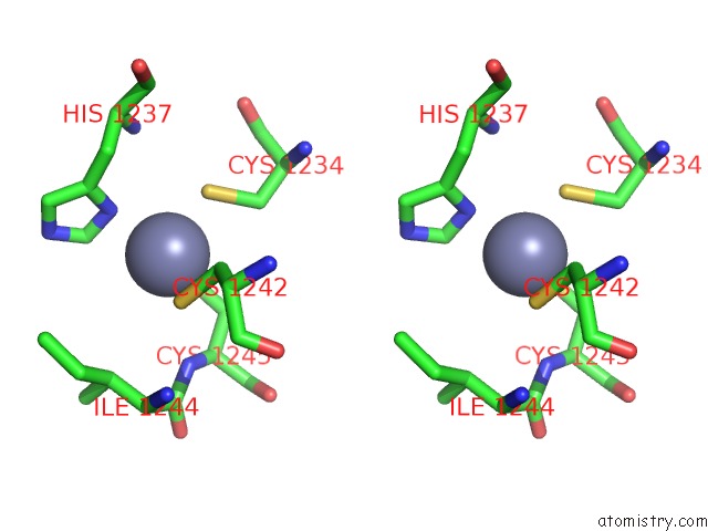
Stereo pair view
Mono view
Stereo pair view
A full contact list of Zinc with other atoms in the Zn binding
site number 2 of Crystal Structure of Tankyrase 1 with Compound 4 within 5.0Å range:
|
Zinc binding site 3 out of 4 in 4i9i
Go back to
Zinc binding site 3 out
of 4 in the Crystal Structure of Tankyrase 1 with Compound 4
Mono view
Stereo pair view
Mono view
Stereo pair view
A full contact list of Zinc with other atoms in the Zn binding
site number 3 of Crystal Structure of Tankyrase 1 with Compound 4 within 5.0Å range:
|
Zinc binding site 4 out of 4 in 4i9i
Go back to
Zinc binding site 4 out
of 4 in the Crystal Structure of Tankyrase 1 with Compound 4
Mono view
Stereo pair view
Mono view
Stereo pair view
A full contact list of Zinc with other atoms in the Zn binding
site number 4 of Crystal Structure of Tankyrase 1 with Compound 4 within 5.0Å range:
|
Reference:
H.Bregman,
H.Gunaydin,
Y.Gu,
S.Schneider,
C.Wilson,
E.F.Dimauro,
X.Huang.
Discovery of A Class of Novel Tankyrase Inhibitors That Bind to Both the Nicotinamide Pocket and the Induced Pocket. J.Med.Chem. V. 56 1341 2013.
ISSN: ISSN 0022-2623
PubMed: 23316926
DOI: 10.1021/JM301607V
Page generated: Wed Aug 20 18:47:40 2025
ISSN: ISSN 0022-2623
PubMed: 23316926
DOI: 10.1021/JM301607V
Last articles
Zn in 5ACSZn in 5ACU
Zn in 5ACT
Zn in 5ACR
Zn in 5ACP
Zn in 5ACQ
Zn in 5ABS
Zn in 5ABX
Zn in 5A7M
Zn in 5ABA