Zinc »
PDB 4hkn-4hwt »
4hlk »
Zinc in PDB 4hlk: Crystal Structure of Tankyrase 2 in Complex with 4'-Methylflavone
Enzymatic activity of Crystal Structure of Tankyrase 2 in Complex with 4'-Methylflavone
All present enzymatic activity of Crystal Structure of Tankyrase 2 in Complex with 4'-Methylflavone:
2.4.2.30;
2.4.2.30;
Protein crystallography data
The structure of Crystal Structure of Tankyrase 2 in Complex with 4'-Methylflavone, PDB code: 4hlk
was solved by
M.Narwal,
T.Haikarainen,
L.Lehtio,
with X-Ray Crystallography technique. A brief refinement statistics is given in the table below:
| Resolution Low / High (Å) | 44.33 / 2.00 |
| Space group | C 2 2 21 |
| Cell size a, b, c (Å), α, β, γ (°) | 93.970, 95.570, 118.250, 90.00, 90.00, 90.00 |
| R / Rfree (%) | 18.4 / 21.5 |
Zinc Binding Sites:
The binding sites of Zinc atom in the Crystal Structure of Tankyrase 2 in Complex with 4'-Methylflavone
(pdb code 4hlk). This binding sites where shown within
5.0 Angstroms radius around Zinc atom.
In total 2 binding sites of Zinc where determined in the Crystal Structure of Tankyrase 2 in Complex with 4'-Methylflavone, PDB code: 4hlk:
Jump to Zinc binding site number: 1; 2;
In total 2 binding sites of Zinc where determined in the Crystal Structure of Tankyrase 2 in Complex with 4'-Methylflavone, PDB code: 4hlk:
Jump to Zinc binding site number: 1; 2;
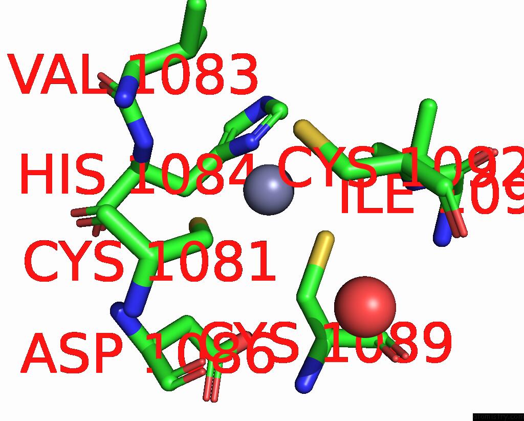
Zinc binding site 1 out of 2 in 4hlk
Go back to
Zinc binding site 1 out
of 2 in the Crystal Structure of Tankyrase 2 in Complex with 4'-Methylflavone
Mono view

Stereo pair view
Mono view
Stereo pair view
A full contact list of Zinc with other atoms in the Zn binding
site number 1 of Crystal Structure of Tankyrase 2 in Complex with 4'-Methylflavone within 5.0Å range:
|
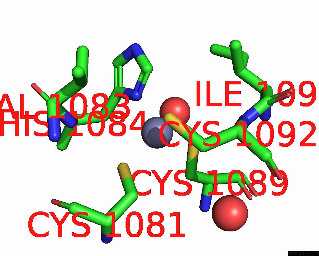
Zinc binding site 2 out of 2 in 4hlk
Go back to
Zinc binding site 2 out
of 2 in the Crystal Structure of Tankyrase 2 in Complex with 4'-Methylflavone
Mono view

Stereo pair view
Mono view
Stereo pair view
A full contact list of Zinc with other atoms in the Zn binding
site number 2 of Crystal Structure of Tankyrase 2 in Complex with 4'-Methylflavone within 5.0Å range:
|
Reference:
M.Narwal,
T.Haikarainen,
A.Fallarero,
P.M.Vuorela,
L.Lehtio.
Screening and Structural Analysis of Flavones Inhibiting Tankyrases. J.Med.Chem. V. 56 3507 2013.
ISSN: ISSN 0022-2623
PubMed: 23574272
DOI: 10.1021/JM3018783
Page generated: Wed Aug 20 18:35:21 2025
ISSN: ISSN 0022-2623
PubMed: 23574272
DOI: 10.1021/JM3018783
Last articles
Zn in 5ADGZn in 5ADF
Zn in 5ADE
Zn in 5ACX
Zn in 5ADD
Zn in 5ACW
Zn in 5ADC
Zn in 5ADB
Zn in 5ADA
Zn in 5AD9