Zinc »
PDB 4gyf-4h8q »
4h54 »
Zinc in PDB 4h54: Crystal Structure of the Diguanylate Cyclase Dgcz
Enzymatic activity of Crystal Structure of the Diguanylate Cyclase Dgcz
All present enzymatic activity of Crystal Structure of the Diguanylate Cyclase Dgcz:
2.7.7.65;
2.7.7.65;
Protein crystallography data
The structure of Crystal Structure of the Diguanylate Cyclase Dgcz, PDB code: 4h54
was solved by
F.Zaehringer,
T.Schirmer,
with X-Ray Crystallography technique. A brief refinement statistics is given in the table below:
| Resolution Low / High (Å) | 73.20 / 3.90 |
| Space group | P 61 |
| Cell size a, b, c (Å), α, β, γ (°) | 102.593, 102.593, 129.156, 90.00, 90.00, 120.00 |
| R / Rfree (%) | 24.2 / 24.1 |
Other elements in 4h54:
The structure of Crystal Structure of the Diguanylate Cyclase Dgcz also contains other interesting chemical elements:
| Magnesium | (Mg) | 2 atoms |
Zinc Binding Sites:
The binding sites of Zinc atom in the Crystal Structure of the Diguanylate Cyclase Dgcz
(pdb code 4h54). This binding sites where shown within
5.0 Angstroms radius around Zinc atom.
In total 2 binding sites of Zinc where determined in the Crystal Structure of the Diguanylate Cyclase Dgcz, PDB code: 4h54:
Jump to Zinc binding site number: 1; 2;
In total 2 binding sites of Zinc where determined in the Crystal Structure of the Diguanylate Cyclase Dgcz, PDB code: 4h54:
Jump to Zinc binding site number: 1; 2;
Zinc binding site 1 out of 2 in 4h54
Go back to
Zinc binding site 1 out
of 2 in the Crystal Structure of the Diguanylate Cyclase Dgcz
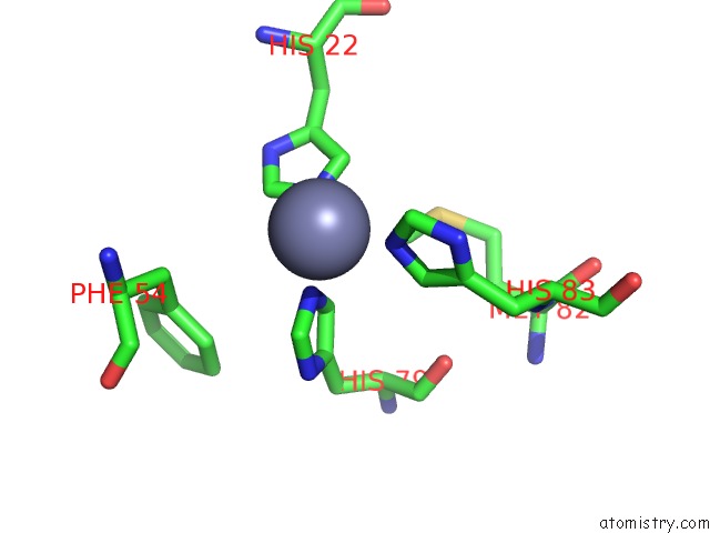
Mono view
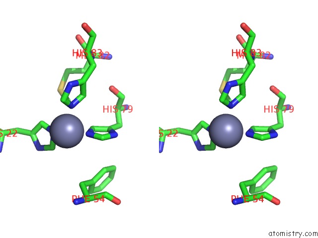
Stereo pair view
Mono view
Stereo pair view
A full contact list of Zinc with other atoms in the Zn binding
site number 1 of Crystal Structure of the Diguanylate Cyclase Dgcz within 5.0Å range:
|
Zinc binding site 2 out of 2 in 4h54
Go back to
Zinc binding site 2 out
of 2 in the Crystal Structure of the Diguanylate Cyclase Dgcz
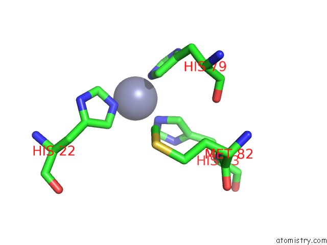
Mono view
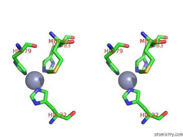
Stereo pair view
Mono view
Stereo pair view
A full contact list of Zinc with other atoms in the Zn binding
site number 2 of Crystal Structure of the Diguanylate Cyclase Dgcz within 5.0Å range:
|
Reference:
F.Zahringer,
E.Lacanna,
U.Jenal,
T.Schirmer,
A.Boehm.
Structure and Signaling Mechanism of A Zinc-Sensory Diguanylate Cyclase. Structure V. 21 1149 2013.
ISSN: ISSN 0969-2126
PubMed: 23769666
DOI: 10.1016/J.STR.2013.04.026
Page generated: Sat Oct 26 23:53:37 2024
ISSN: ISSN 0969-2126
PubMed: 23769666
DOI: 10.1016/J.STR.2013.04.026
Last articles
Zn in 9MJ5Zn in 9HNW
Zn in 9G0L
Zn in 9FNE
Zn in 9DZN
Zn in 9E0I
Zn in 9D32
Zn in 9DAK
Zn in 8ZXC
Zn in 8ZUF