Zinc »
PDB 4gyf-4h8q »
4h3x »
Zinc in PDB 4h3x: Crystal Structure of An Mmp Broad Spectrum Hydroxamate Based Inhibitor CC27 in Complex with the Mmp-9 Catalytic Domain
Enzymatic activity of Crystal Structure of An Mmp Broad Spectrum Hydroxamate Based Inhibitor CC27 in Complex with the Mmp-9 Catalytic Domain
All present enzymatic activity of Crystal Structure of An Mmp Broad Spectrum Hydroxamate Based Inhibitor CC27 in Complex with the Mmp-9 Catalytic Domain:
3.4.24.35;
3.4.24.35;
Protein crystallography data
The structure of Crystal Structure of An Mmp Broad Spectrum Hydroxamate Based Inhibitor CC27 in Complex with the Mmp-9 Catalytic Domain, PDB code: 4h3x
was solved by
E.A.Stura,
L.Vera,
E.Cassar-Lajeunesse,
E.Nuti,
V.Dive,
A.Rossello,
with X-Ray Crystallography technique. A brief refinement statistics is given in the table below:
| Resolution Low / High (Å) | 42.81 / 1.76 |
| Space group | P 1 21 1 |
| Cell size a, b, c (Å), α, β, γ (°) | 40.110, 97.940, 46.080, 90.00, 111.73, 90.00 |
| R / Rfree (%) | 19.6 / 25.4 |
Other elements in 4h3x:
The structure of Crystal Structure of An Mmp Broad Spectrum Hydroxamate Based Inhibitor CC27 in Complex with the Mmp-9 Catalytic Domain also contains other interesting chemical elements:
| Calcium | (Ca) | 6 atoms |
Zinc Binding Sites:
The binding sites of Zinc atom in the Crystal Structure of An Mmp Broad Spectrum Hydroxamate Based Inhibitor CC27 in Complex with the Mmp-9 Catalytic Domain
(pdb code 4h3x). This binding sites where shown within
5.0 Angstroms radius around Zinc atom.
In total 4 binding sites of Zinc where determined in the Crystal Structure of An Mmp Broad Spectrum Hydroxamate Based Inhibitor CC27 in Complex with the Mmp-9 Catalytic Domain, PDB code: 4h3x:
Jump to Zinc binding site number: 1; 2; 3; 4;
In total 4 binding sites of Zinc where determined in the Crystal Structure of An Mmp Broad Spectrum Hydroxamate Based Inhibitor CC27 in Complex with the Mmp-9 Catalytic Domain, PDB code: 4h3x:
Jump to Zinc binding site number: 1; 2; 3; 4;
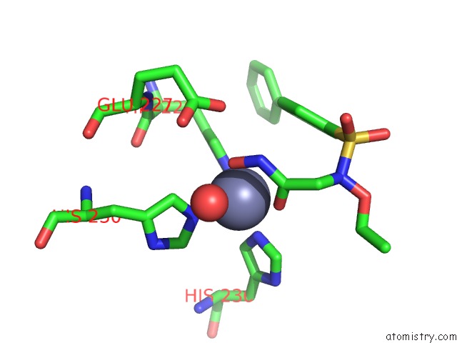
Zinc binding site 1 out of 4 in 4h3x
Go back to
Zinc binding site 1 out
of 4 in the Crystal Structure of An Mmp Broad Spectrum Hydroxamate Based Inhibitor CC27 in Complex with the Mmp-9 Catalytic Domain
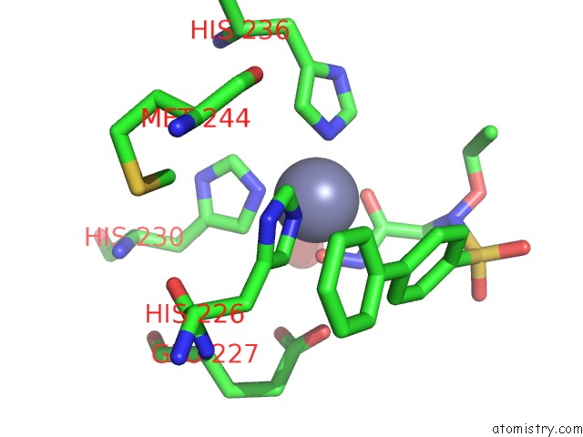
Mono view
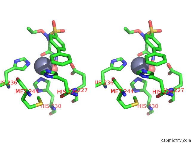
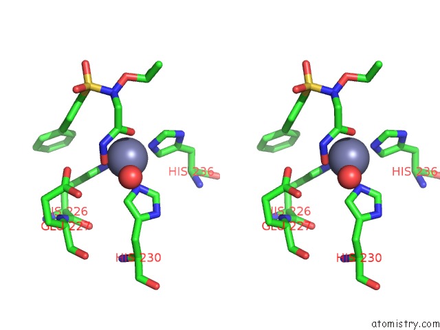
Stereo pair view
Mono view
Stereo pair view
A full contact list of Zinc with other atoms in the Zn binding
site number 1 of Crystal Structure of An Mmp Broad Spectrum Hydroxamate Based Inhibitor CC27 in Complex with the Mmp-9 Catalytic Domain within 5.0Å range:
|
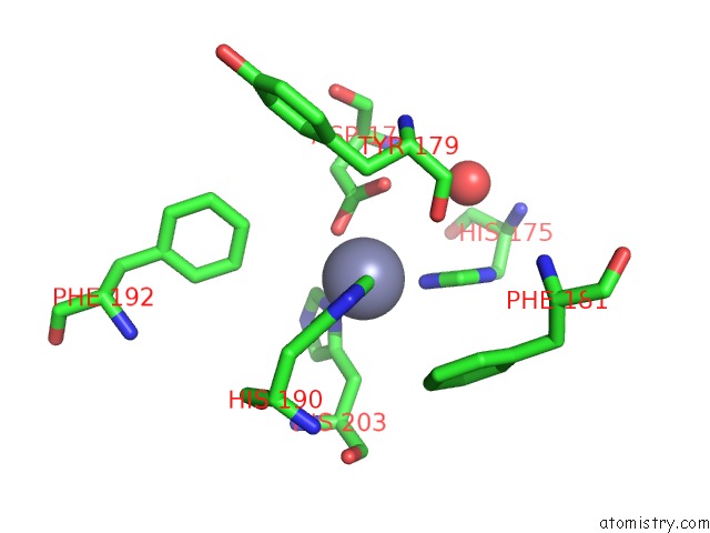
Zinc binding site 2 out of 4 in 4h3x
Go back to
Zinc binding site 2 out
of 4 in the Crystal Structure of An Mmp Broad Spectrum Hydroxamate Based Inhibitor CC27 in Complex with the Mmp-9 Catalytic Domain
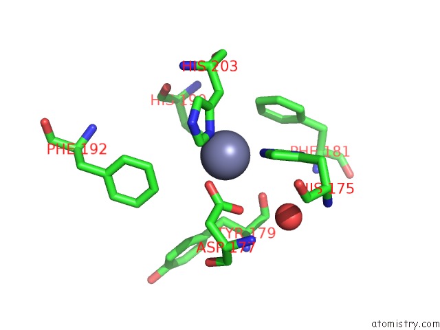
Mono view
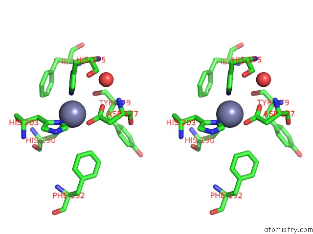
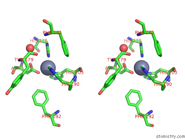
Stereo pair view
Mono view
Stereo pair view
A full contact list of Zinc with other atoms in the Zn binding
site number 2 of Crystal Structure of An Mmp Broad Spectrum Hydroxamate Based Inhibitor CC27 in Complex with the Mmp-9 Catalytic Domain within 5.0Å range:
|
Zinc binding site 3 out of 4 in 4h3x
Go back to
Zinc binding site 3 out
of 4 in the Crystal Structure of An Mmp Broad Spectrum Hydroxamate Based Inhibitor CC27 in Complex with the Mmp-9 Catalytic Domain
Mono view
Stereo pair view
Mono view
Stereo pair view
A full contact list of Zinc with other atoms in the Zn binding
site number 3 of Crystal Structure of An Mmp Broad Spectrum Hydroxamate Based Inhibitor CC27 in Complex with the Mmp-9 Catalytic Domain within 5.0Å range:
|
Zinc binding site 4 out of 4 in 4h3x
Go back to
Zinc binding site 4 out
of 4 in the Crystal Structure of An Mmp Broad Spectrum Hydroxamate Based Inhibitor CC27 in Complex with the Mmp-9 Catalytic Domain
Mono view
Stereo pair view
Mono view
Stereo pair view
A full contact list of Zinc with other atoms in the Zn binding
site number 4 of Crystal Structure of An Mmp Broad Spectrum Hydroxamate Based Inhibitor CC27 in Complex with the Mmp-9 Catalytic Domain within 5.0Å range:
|
Reference:
C.Antoni,
L.Vera,
L.Devel,
M.P.Catalani,
B.Czarny,
E.Cassar-Lajeunesse,
E.Nuti,
A.Rossello,
V.Dive,
E.A.Stura.
Crystallization of Bi-Functional Ligand Protein Complexes. J.Struct.Biol. V. 182 246 2013.
ISSN: ISSN 1047-8477
PubMed: 23567804
DOI: 10.1016/J.JSB.2013.03.015
Page generated: Sat Oct 26 23:52:47 2024
ISSN: ISSN 1047-8477
PubMed: 23567804
DOI: 10.1016/J.JSB.2013.03.015
Last articles
Zn in 9MJ5Zn in 9HNW
Zn in 9G0L
Zn in 9FNE
Zn in 9DZN
Zn in 9E0I
Zn in 9D32
Zn in 9DAK
Zn in 8ZXC
Zn in 8ZUF