Zinc »
PDB 4ggk-4gsz »
4giz »
Zinc in PDB 4giz: Crystal Structure of Full-Length Human Papillomavirus Oncoprotein E6 in Complex with Lxxll Peptide of Ubiquitin Ligase E6AP at 2.55 A Resolution
Protein crystallography data
The structure of Crystal Structure of Full-Length Human Papillomavirus Oncoprotein E6 in Complex with Lxxll Peptide of Ubiquitin Ligase E6AP at 2.55 A Resolution, PDB code: 4giz
was solved by
A.G.Mcewen,
K.Zanier,
S.Charbonnier,
P.Poussin,
V.Cura,
S.Vande Pol,
G.Trave,
J.Cavarelli,
with X-Ray Crystallography technique. A brief refinement statistics is given in the table below:
| Resolution Low / High (Å) | 37.33 / 2.55 |
| Space group | P 21 21 21 |
| Cell size a, b, c (Å), α, β, γ (°) | 106.279, 134.936, 138.794, 90.00, 90.00, 90.00 |
| R / Rfree (%) | 16.6 / 19.6 |
Zinc Binding Sites:
The binding sites of Zinc atom in the Crystal Structure of Full-Length Human Papillomavirus Oncoprotein E6 in Complex with Lxxll Peptide of Ubiquitin Ligase E6AP at 2.55 A Resolution
(pdb code 4giz). This binding sites where shown within
5.0 Angstroms radius around Zinc atom.
In total 4 binding sites of Zinc where determined in the Crystal Structure of Full-Length Human Papillomavirus Oncoprotein E6 in Complex with Lxxll Peptide of Ubiquitin Ligase E6AP at 2.55 A Resolution, PDB code: 4giz:
Jump to Zinc binding site number: 1; 2; 3; 4;
In total 4 binding sites of Zinc where determined in the Crystal Structure of Full-Length Human Papillomavirus Oncoprotein E6 in Complex with Lxxll Peptide of Ubiquitin Ligase E6AP at 2.55 A Resolution, PDB code: 4giz:
Jump to Zinc binding site number: 1; 2; 3; 4;
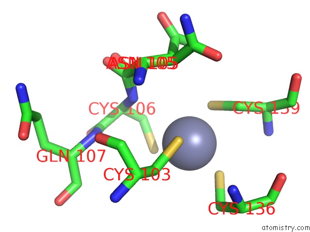
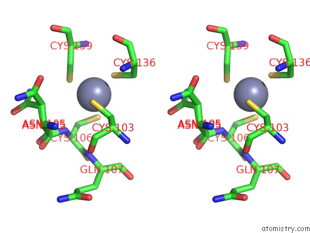
Zinc binding site 1 out of 4 in 4giz
Go back to
Zinc binding site 1 out
of 4 in the Crystal Structure of Full-Length Human Papillomavirus Oncoprotein E6 in Complex with Lxxll Peptide of Ubiquitin Ligase E6AP at 2.55 A Resolution
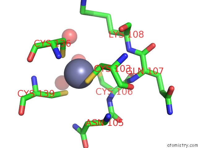
Mono view
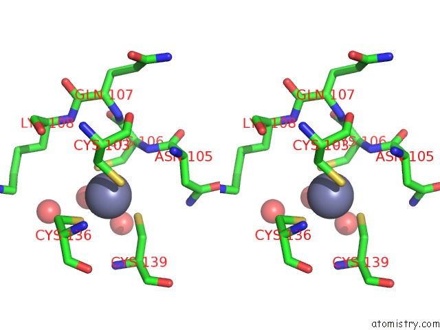
Stereo pair view
Mono view
Stereo pair view
A full contact list of Zinc with other atoms in the Zn binding
site number 1 of Crystal Structure of Full-Length Human Papillomavirus Oncoprotein E6 in Complex with Lxxll Peptide of Ubiquitin Ligase E6AP at 2.55 A Resolution within 5.0Å range:
|
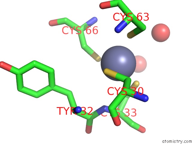
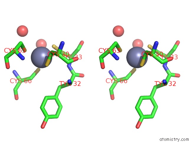
Zinc binding site 2 out of 4 in 4giz
Go back to
Zinc binding site 2 out
of 4 in the Crystal Structure of Full-Length Human Papillomavirus Oncoprotein E6 in Complex with Lxxll Peptide of Ubiquitin Ligase E6AP at 2.55 A Resolution
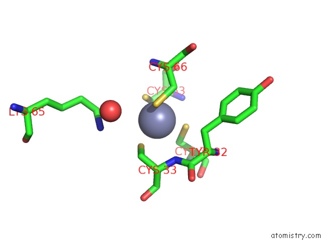
Mono view
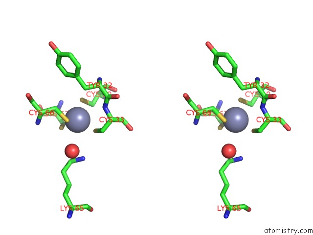
Stereo pair view
Mono view
Stereo pair view
A full contact list of Zinc with other atoms in the Zn binding
site number 2 of Crystal Structure of Full-Length Human Papillomavirus Oncoprotein E6 in Complex with Lxxll Peptide of Ubiquitin Ligase E6AP at 2.55 A Resolution within 5.0Å range:
|
Zinc binding site 3 out of 4 in 4giz
Go back to
Zinc binding site 3 out
of 4 in the Crystal Structure of Full-Length Human Papillomavirus Oncoprotein E6 in Complex with Lxxll Peptide of Ubiquitin Ligase E6AP at 2.55 A Resolution
Mono view
Stereo pair view
Mono view
Stereo pair view
A full contact list of Zinc with other atoms in the Zn binding
site number 3 of Crystal Structure of Full-Length Human Papillomavirus Oncoprotein E6 in Complex with Lxxll Peptide of Ubiquitin Ligase E6AP at 2.55 A Resolution within 5.0Å range:
|
Zinc binding site 4 out of 4 in 4giz
Go back to
Zinc binding site 4 out
of 4 in the Crystal Structure of Full-Length Human Papillomavirus Oncoprotein E6 in Complex with Lxxll Peptide of Ubiquitin Ligase E6AP at 2.55 A Resolution
Mono view
Stereo pair view
Mono view
Stereo pair view
A full contact list of Zinc with other atoms in the Zn binding
site number 4 of Crystal Structure of Full-Length Human Papillomavirus Oncoprotein E6 in Complex with Lxxll Peptide of Ubiquitin Ligase E6AP at 2.55 A Resolution within 5.0Å range:
|
Reference:
K.Zanier,
S.Charbonnier,
A.O.Sidi,
A.G.Mcewen,
M.G.Ferrario,
P.Poussin-Courmontagne,
V.Cura,
N.Brimer,
K.O.Babah,
T.Ansari,
I.Muller,
R.H.Stote,
J.Cavarelli,
S.Vande Pol,
G.Trave.
Structural Basis For Hijacking of Cellular Lxxll Motifs By Papillomavirus E6 Oncoproteins. Science V. 339 694 2013.
ISSN: ISSN 0036-8075
PubMed: 23393263
DOI: 10.1126/SCIENCE.1229934
Page generated: Sat Oct 26 23:19:31 2024
ISSN: ISSN 0036-8075
PubMed: 23393263
DOI: 10.1126/SCIENCE.1229934
Last articles
Zn in 9MJ5Zn in 9HNW
Zn in 9G0L
Zn in 9FNE
Zn in 9DZN
Zn in 9E0I
Zn in 9D32
Zn in 9DAK
Zn in 8ZXC
Zn in 8ZUF