Zinc »
PDB 4g3m-4ggj »
4gbd »
Zinc in PDB 4gbd: Crystal Structure of Adenosine Deaminase From Pseudomonas Aeruginosa PAO1 with Bound Zn and Methylthio-Coformycin
Protein crystallography data
The structure of Crystal Structure of Adenosine Deaminase From Pseudomonas Aeruginosa PAO1 with Bound Zn and Methylthio-Coformycin, PDB code: 4gbd
was solved by
M.Ho,
R.Guan,
S.C.Almo,
V.L.Schramm,
with X-Ray Crystallography technique. A brief refinement statistics is given in the table below:
| Resolution Low / High (Å) | 31.10 / 1.98 |
| Space group | C 1 2 1 |
| Cell size a, b, c (Å), α, β, γ (°) | 119.814, 120.310, 77.298, 90.00, 108.00, 90.00 |
| R / Rfree (%) | 19 / 23.1 |
Zinc Binding Sites:
The binding sites of Zinc atom in the Crystal Structure of Adenosine Deaminase From Pseudomonas Aeruginosa PAO1 with Bound Zn and Methylthio-Coformycin
(pdb code 4gbd). This binding sites where shown within
5.0 Angstroms radius around Zinc atom.
In total 4 binding sites of Zinc where determined in the Crystal Structure of Adenosine Deaminase From Pseudomonas Aeruginosa PAO1 with Bound Zn and Methylthio-Coformycin, PDB code: 4gbd:
Jump to Zinc binding site number: 1; 2; 3; 4;
In total 4 binding sites of Zinc where determined in the Crystal Structure of Adenosine Deaminase From Pseudomonas Aeruginosa PAO1 with Bound Zn and Methylthio-Coformycin, PDB code: 4gbd:
Jump to Zinc binding site number: 1; 2; 3; 4;
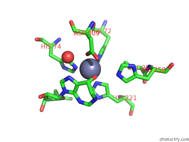
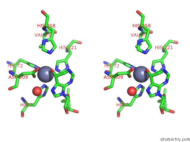
Zinc binding site 1 out of 4 in 4gbd
Go back to
Zinc binding site 1 out
of 4 in the Crystal Structure of Adenosine Deaminase From Pseudomonas Aeruginosa PAO1 with Bound Zn and Methylthio-Coformycin
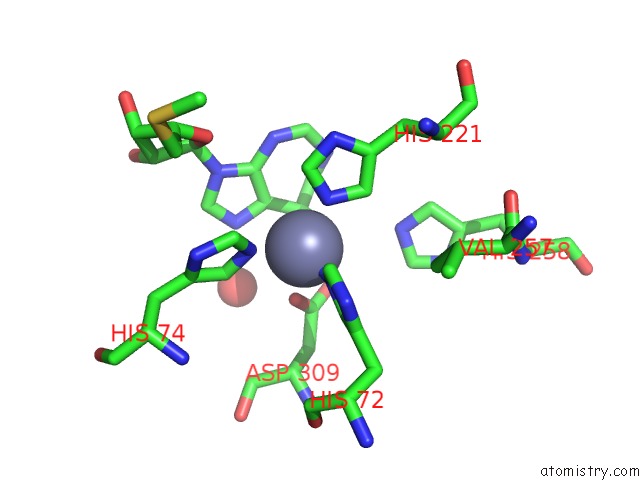
Mono view
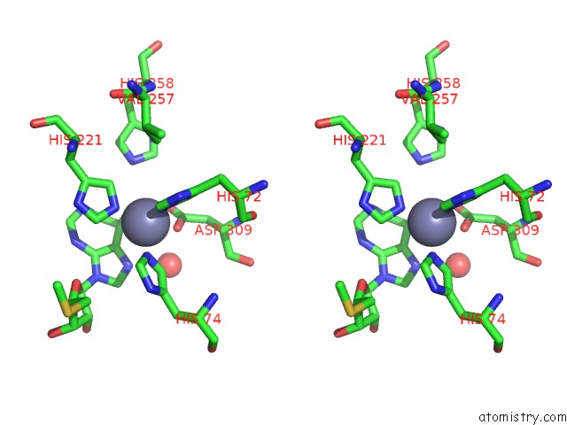
Stereo pair view
Mono view
Stereo pair view
A full contact list of Zinc with other atoms in the Zn binding
site number 1 of Crystal Structure of Adenosine Deaminase From Pseudomonas Aeruginosa PAO1 with Bound Zn and Methylthio-Coformycin within 5.0Å range:
|
Zinc binding site 2 out of 4 in 4gbd
Go back to
Zinc binding site 2 out
of 4 in the Crystal Structure of Adenosine Deaminase From Pseudomonas Aeruginosa PAO1 with Bound Zn and Methylthio-Coformycin
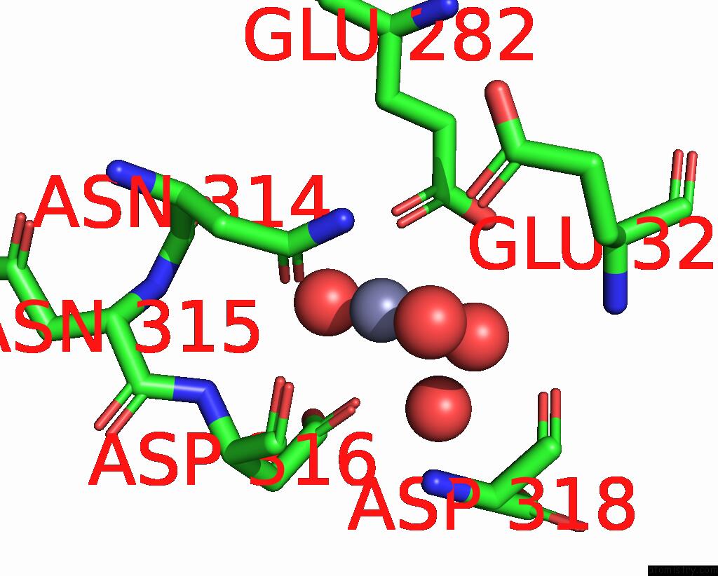
Mono view
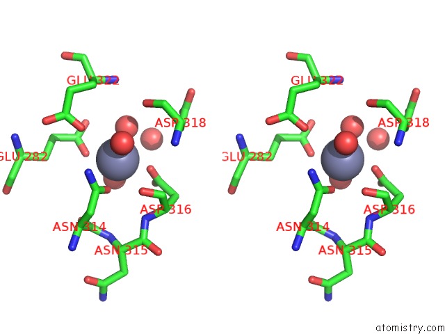
Stereo pair view
Mono view
Stereo pair view
A full contact list of Zinc with other atoms in the Zn binding
site number 2 of Crystal Structure of Adenosine Deaminase From Pseudomonas Aeruginosa PAO1 with Bound Zn and Methylthio-Coformycin within 5.0Å range:
|
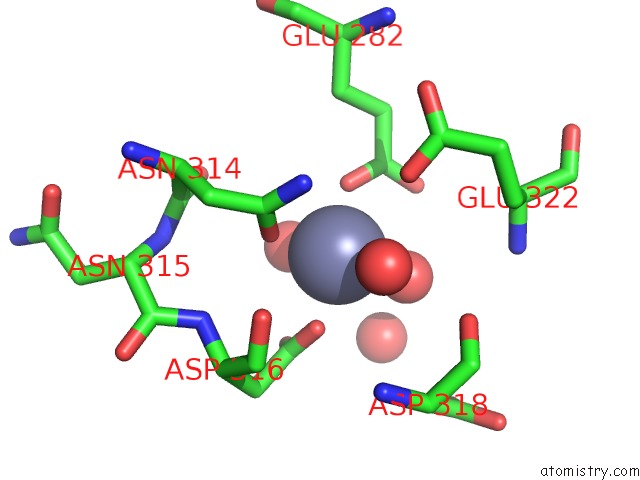
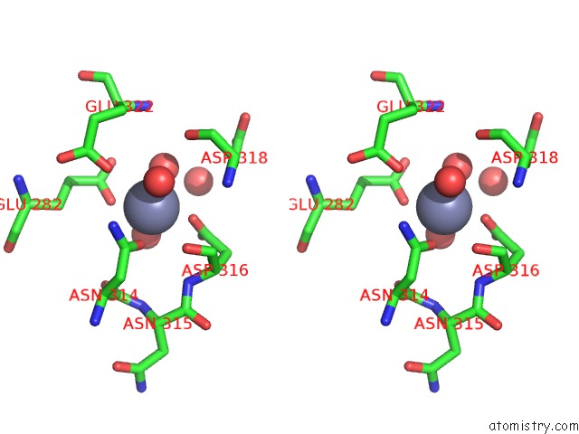
Zinc binding site 3 out of 4 in 4gbd
Go back to
Zinc binding site 3 out
of 4 in the Crystal Structure of Adenosine Deaminase From Pseudomonas Aeruginosa PAO1 with Bound Zn and Methylthio-Coformycin
Mono view
Stereo pair view
Mono view
Stereo pair view
A full contact list of Zinc with other atoms in the Zn binding
site number 3 of Crystal Structure of Adenosine Deaminase From Pseudomonas Aeruginosa PAO1 with Bound Zn and Methylthio-Coformycin within 5.0Å range:
|
Zinc binding site 4 out of 4 in 4gbd
Go back to
Zinc binding site 4 out
of 4 in the Crystal Structure of Adenosine Deaminase From Pseudomonas Aeruginosa PAO1 with Bound Zn and Methylthio-Coformycin
Mono view
Stereo pair view
Mono view
Stereo pair view
A full contact list of Zinc with other atoms in the Zn binding
site number 4 of Crystal Structure of Adenosine Deaminase From Pseudomonas Aeruginosa PAO1 with Bound Zn and Methylthio-Coformycin within 5.0Å range:
|
Reference:
R.Guan,
M.C.Ho,
R.F.Frohlich,
P.C.Tyler,
S.C.Almo,
V.L.Schramm.
Methylthioadenosine Deaminase in An Alternative Quorum Sensing Pathway in Pseudomonas Aeruginosa. Biochemistry V. 51 9094 2012.
ISSN: ISSN 0006-2960
PubMed: 23050701
DOI: 10.1021/BI301062Y
Page generated: Sat Oct 26 23:12:30 2024
ISSN: ISSN 0006-2960
PubMed: 23050701
DOI: 10.1021/BI301062Y
Last articles
As in 3NLTAs in 3NRF
As in 3NLI
As in 3NLG
As in 3NLH
As in 3NLF
As in 3N6G
As in 3NLE
As in 3NLD
As in 3N6F