Zinc »
PDB 4g3m-4ggj »
4gbc »
Zinc in PDB 4gbc: Crystal Structure of Aspart Insulin at pH 6.5
Protein crystallography data
The structure of Crystal Structure of Aspart Insulin at pH 6.5, PDB code: 4gbc
was solved by
L.M.T.R.Lima,
M.P.Favero-Retto,
L.C.Palmieri,
with X-Ray Crystallography technique. A brief refinement statistics is given in the table below:
| Resolution Low / High (Å) | 39.08 / 1.78 |
| Space group | H 3 |
| Cell size a, b, c (Å), α, β, γ (°) | 78.150, 78.150, 36.850, 90.00, 90.00, 120.00 |
| R / Rfree (%) | 16.7 / 22.4 |
Other elements in 4gbc:
The structure of Crystal Structure of Aspart Insulin at pH 6.5 also contains other interesting chemical elements:
| Chlorine | (Cl) | 2 atoms |
Zinc Binding Sites:
The binding sites of Zinc atom in the Crystal Structure of Aspart Insulin at pH 6.5
(pdb code 4gbc). This binding sites where shown within
5.0 Angstroms radius around Zinc atom.
In total 2 binding sites of Zinc where determined in the Crystal Structure of Aspart Insulin at pH 6.5, PDB code: 4gbc:
Jump to Zinc binding site number: 1; 2;
In total 2 binding sites of Zinc where determined in the Crystal Structure of Aspart Insulin at pH 6.5, PDB code: 4gbc:
Jump to Zinc binding site number: 1; 2;
Zinc binding site 1 out of 2 in 4gbc
Go back to
Zinc binding site 1 out
of 2 in the Crystal Structure of Aspart Insulin at pH 6.5
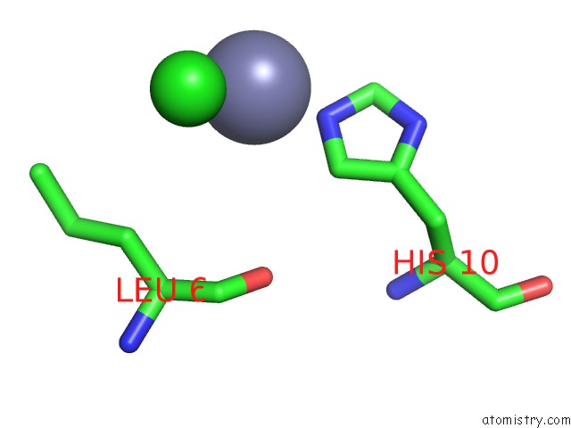
Mono view
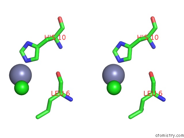
Stereo pair view
Mono view
Stereo pair view
A full contact list of Zinc with other atoms in the Zn binding
site number 1 of Crystal Structure of Aspart Insulin at pH 6.5 within 5.0Å range:
|
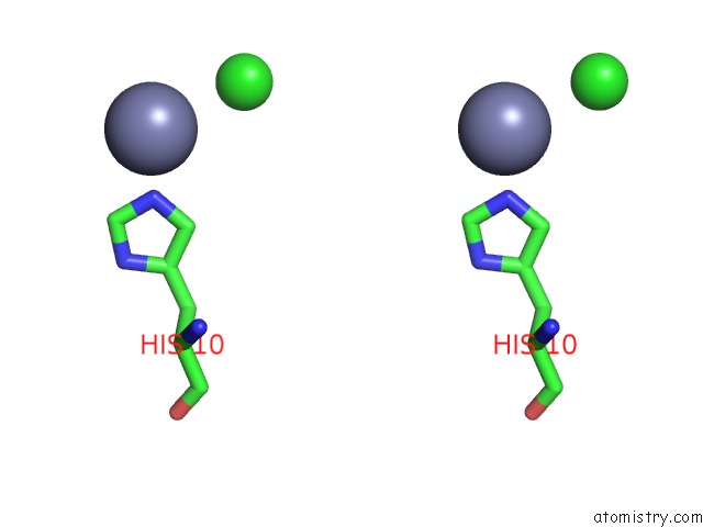
Zinc binding site 2 out of 2 in 4gbc
Go back to
Zinc binding site 2 out
of 2 in the Crystal Structure of Aspart Insulin at pH 6.5
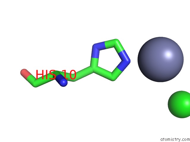
Mono view
Stereo pair view
Mono view
Stereo pair view
A full contact list of Zinc with other atoms in the Zn binding
site number 2 of Crystal Structure of Aspart Insulin at pH 6.5 within 5.0Å range:
|
Reference:
L.C.Palmieri,
M.P.Favero-Retto,
D.Lourenco,
L.M.Lima.
A T3R3 Hexamer of the Human Insulin Variant B28ASP. Biophys.Chem. V. 173 1 2013.
ISSN: ISSN 0301-4622
PubMed: 23428413
DOI: 10.1016/J.BPC.2013.01.003
Page generated: Sat Oct 26 23:12:30 2024
ISSN: ISSN 0301-4622
PubMed: 23428413
DOI: 10.1016/J.BPC.2013.01.003
Last articles
Zn in 9MJ5Zn in 9HNW
Zn in 9G0L
Zn in 9FNE
Zn in 9DZN
Zn in 9E0I
Zn in 9D32
Zn in 9DAK
Zn in 8ZXC
Zn in 8ZUF