Zinc in PDB 4f52: Structure of A Glomulin-RBX1-CUL1 Complex
Protein crystallography data
The structure of Structure of A Glomulin-RBX1-CUL1 Complex, PDB code: 4f52
was solved by
D.M.Duda,
J.L.Olszewski,
B.A.Schulman,
with X-Ray Crystallography technique. A brief refinement statistics is given in the table below:
Resolution Low / High (Å)
|
37.39 /
3.00
|
Space group
|
P 1 21 1
|
Cell size a, b, c (Å), α, β, γ (°)
|
53.333,
193.932,
142.075,
90.00,
98.81,
90.00
|
R / Rfree (%)
|
21.9 /
28.9
|
Zinc Binding Sites:
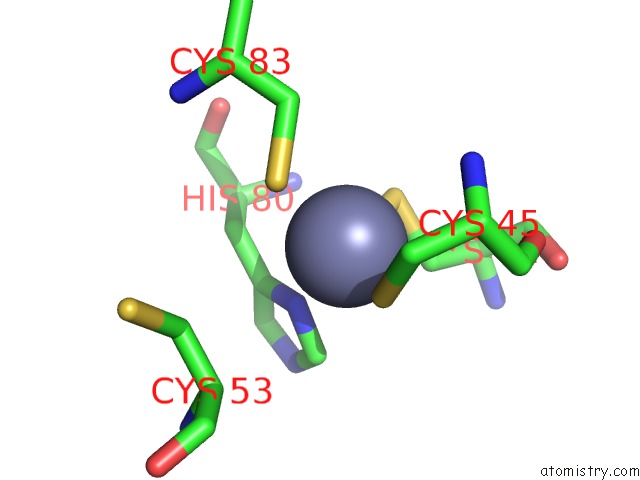
The binding sites of Zinc atom in the Structure of A Glomulin-RBX1-CUL1 Complex
(pdb code 4f52). This binding sites where shown within
5.0 Angstroms radius around Zinc atom.
In total 6 binding sites of Zinc where determined in the
Structure of A Glomulin-RBX1-CUL1 Complex, PDB code: 4f52:
Jump to Zinc binding site number:
1;
2;
3;
4;
5;
6;
Zinc binding site 1 out
of 6 in 4f52
Go back to
Zinc Binding Sites List in 4f52
Zinc binding site 1 out
of 6 in the Structure of A Glomulin-RBX1-CUL1 Complex
Mono view
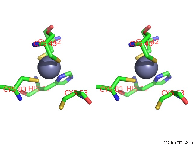
Stereo pair view
|
A full contact list of Zinc with other atoms in the Zn binding
site number 1 of Structure of A Glomulin-RBX1-CUL1 Complex within 5.0Å range:
probe
|
atom
|
residue
|
distance (Å)
|
B
|
Occ
|
B:Zn201
b:91.5
occ:1.00
|
ND1
|
B:HIS80
|
2.1
|
0.0
|
1.0
|
SG
|
B:CYS42
|
2.1
|
88.3
|
1.0
|
SG
|
B:CYS45
|
2.3
|
0.4
|
1.0
|
SG
|
B:CYS83
|
2.4
|
84.1
|
1.0
|
CB
|
B:CYS42
|
2.7
|
88.8
|
1.0
|
CG
|
B:HIS80
|
3.0
|
0.3
|
1.0
|
CE1
|
B:HIS80
|
3.0
|
0.4
|
1.0
|
CB
|
B:HIS80
|
3.3
|
0.0
|
1.0
|
CB
|
B:CYS45
|
3.5
|
0.4
|
1.0
|
CB
|
B:CYS83
|
3.6
|
0.8
|
1.0
|
N
|
B:CYS45
|
3.9
|
0.1
|
1.0
|
NE2
|
B:HIS80
|
4.0
|
0.7
|
1.0
|
CD2
|
B:HIS80
|
4.1
|
0.8
|
1.0
|
CA
|
B:CYS42
|
4.2
|
83.3
|
1.0
|
CA
|
B:CYS45
|
4.2
|
0.5
|
1.0
|
CB
|
B:CYS53
|
4.2
|
93.5
|
1.0
|
N
|
B:HIS80
|
4.2
|
97.8
|
1.0
|
O
|
B:CYS45
|
4.2
|
0.9
|
1.0
|
C
|
B:CYS45
|
4.3
|
0.8
|
1.0
|
CA
|
B:HIS80
|
4.4
|
0.2
|
1.0
|
CA
|
B:CYS53
|
4.4
|
0.9
|
1.0
|
C
|
B:CYS42
|
4.7
|
74.4
|
1.0
|
CA
|
B:CYS83
|
4.9
|
0.9
|
1.0
|
O
|
B:CYS42
|
5.0
|
67.1
|
1.0
|
|
Zinc binding site 2 out
of 6 in 4f52
Go back to
Zinc Binding Sites List in 4f52
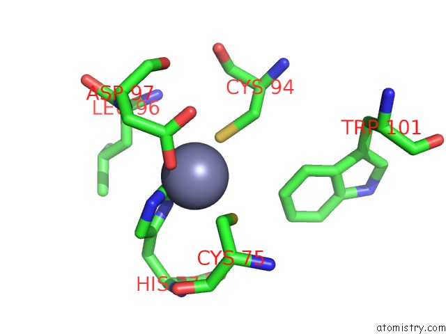
Zinc binding site 2 out
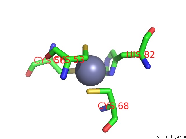
of 6 in the Structure of A Glomulin-RBX1-CUL1 Complex
Mono view
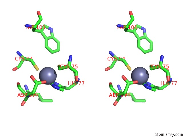
Stereo pair view
|
A full contact list of Zinc with other atoms in the Zn binding
site number 2 of Structure of A Glomulin-RBX1-CUL1 Complex within 5.0Å range:
probe
|
atom
|
residue
|
distance (Å)
|
B
|
Occ
|
B:Zn202
b:96.8
occ:1.00
|
CE1
|
B:HIS77
|
1.2
|
0.1
|
1.0
|
ND1
|
B:HIS77
|
2.1
|
0.8
|
1.0
|
OD2
|
B:ASP97
|
2.1
|
0.9
|
1.0
|
NE2
|
B:HIS77
|
2.2
|
0.4
|
1.0
|
SG
|
B:CYS94
|
2.3
|
96.2
|
1.0
|
SG
|
B:CYS75
|
2.4
|
0.8
|
1.0
|
CG
|
B:ASP97
|
3.0
|
0.7
|
1.0
|
CB
|
B:CYS75
|
3.1
|
0.8
|
1.0
|
CG
|
B:HIS77
|
3.1
|
0.6
|
1.0
|
CD2
|
B:HIS77
|
3.2
|
0.3
|
1.0
|
OD1
|
B:ASP97
|
3.4
|
0.8
|
1.0
|
CB
|
B:CYS94
|
3.6
|
96.7
|
1.0
|
N
|
B:ASP97
|
4.0
|
0.4
|
1.0
|
CB
|
B:ASP97
|
4.1
|
1.0
|
1.0
|
CZ3
|
B:TRP101
|
4.2
|
0.2
|
1.0
|
CB
|
B:LEU96
|
4.2
|
0.6
|
1.0
|
CB
|
B:HIS77
|
4.5
|
0.8
|
1.0
|
CA
|
B:CYS75
|
4.5
|
0.5
|
1.0
|
CA
|
B:ASP97
|
4.6
|
0.7
|
1.0
|
N
|
B:LEU96
|
4.7
|
0.4
|
1.0
|
O
|
B:ASP97
|
4.8
|
0.1
|
1.0
|
CE3
|
B:TRP101
|
4.8
|
0.0
|
1.0
|
CA
|
B:LEU96
|
4.8
|
0.9
|
1.0
|
N
|
B:HIS77
|
4.9
|
0.6
|
1.0
|
C
|
B:CYS75
|
4.9
|
0.5
|
1.0
|
CA
|
B:CYS94
|
4.9
|
0.1
|
1.0
|
C
|
B:LEU96
|
4.9
|
0.1
|
1.0
|
C
|
B:ASP97
|
4.9
|
0.7
|
1.0
|
|
Zinc binding site 3 out
of 6 in 4f52
Go back to
Zinc Binding Sites List in 4f52
Zinc binding site 3 out
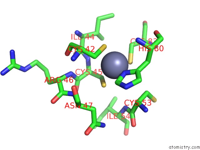
of 6 in the Structure of A Glomulin-RBX1-CUL1 Complex
Mono view
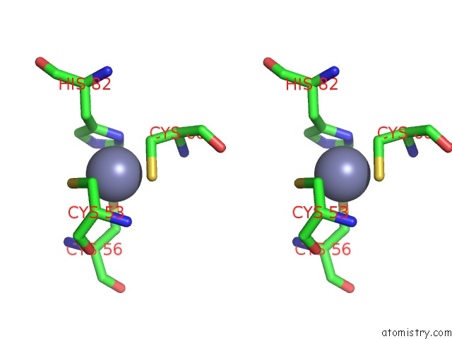
Stereo pair view
|
A full contact list of Zinc with other atoms in the Zn binding
site number 3 of Structure of A Glomulin-RBX1-CUL1 Complex within 5.0Å range:
probe
|
atom
|
residue
|
distance (Å)
|
B
|
Occ
|
B:Zn203
b:0.3
occ:1.00
|
ND1
|
B:HIS82
|
2.1
|
0.2
|
1.0
|
SG
|
B:CYS56
|
2.3
|
0.2
|
1.0
|
SG
|
B:CYS68
|
2.3
|
86.8
|
1.0
|
SG
|
B:CYS53
|
2.6
|
0.1
|
1.0
|
CB
|
B:CYS68
|
2.6
|
0.8
|
1.0
|
CG
|
B:HIS82
|
2.6
|
0.2
|
1.0
|
CE1
|
B:HIS82
|
2.6
|
0.8
|
1.0
|
CB
|
B:CYS56
|
2.9
|
0.7
|
1.0
|
CD2
|
B:HIS82
|
3.2
|
0.7
|
1.0
|
NE2
|
B:HIS82
|
3.2
|
0.9
|
1.0
|
CB
|
B:HIS82
|
3.3
|
0.7
|
1.0
|
CB
|
B:CYS53
|
3.3
|
93.5
|
1.0
|
CA
|
B:CYS68
|
3.7
|
0.1
|
1.0
|
N
|
B:CYS68
|
3.7
|
0.3
|
1.0
|
CA
|
B:CYS56
|
4.1
|
0.4
|
1.0
|
N
|
B:CYS56
|
4.1
|
0.2
|
1.0
|
CA
|
B:CYS53
|
4.3
|
0.9
|
1.0
|
O
|
B:CYS53
|
4.4
|
0.1
|
1.0
|
N
|
B:CYS53
|
4.4
|
0.4
|
1.0
|
C
|
B:CYS53
|
4.6
|
0.1
|
1.0
|
CA
|
B:HIS82
|
4.7
|
0.7
|
1.0
|
C
|
B:CYS68
|
4.9
|
0.1
|
1.0
|
|
Zinc binding site 4 out
of 6 in 4f52
Go back to
Zinc Binding Sites List in 4f52
Zinc binding site 4 out
of 6 in the Structure of A Glomulin-RBX1-CUL1 Complex
Mono view
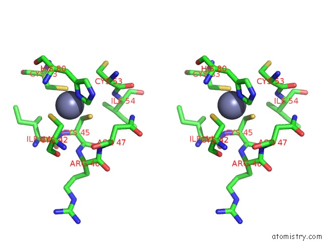
Stereo pair view
|
A full contact list of Zinc with other atoms in the Zn binding
site number 4 of Structure of A Glomulin-RBX1-CUL1 Complex within 5.0Å range:
probe
|
atom
|
residue
|
distance (Å)
|
B
|
Occ
|
D:Zn201
b:79.1
occ:1.00
|
ND1
|
D:HIS80
|
2.1
|
90.0
|
1.0
|
SG
|
D:CYS42
|
2.2
|
0.8
|
1.0
|
SG
|
D:CYS45
|
2.4
|
88.9
|
1.0
|
SG
|
D:CYS83
|
2.5
|
89.5
|
1.0
|
CB
|
D:CYS42
|
2.9
|
68.9
|
1.0
|
CE1
|
D:HIS80
|
2.9
|
90.7
|
1.0
|
CB
|
D:CYS45
|
3.0
|
83.8
|
1.0
|
CG
|
D:HIS80
|
3.1
|
84.5
|
1.0
|
CB
|
D:CYS83
|
3.5
|
0.2
|
1.0
|
CB
|
D:HIS80
|
3.6
|
83.1
|
1.0
|
N
|
D:CYS45
|
3.6
|
81.8
|
1.0
|
CA
|
D:CYS45
|
3.8
|
78.3
|
1.0
|
NE2
|
D:HIS80
|
4.1
|
84.9
|
1.0
|
N
|
D:HIS80
|
4.2
|
83.2
|
1.0
|
CD2
|
D:HIS80
|
4.2
|
81.1
|
1.0
|
CA
|
D:CYS42
|
4.4
|
73.0
|
1.0
|
CB
|
D:CYS53
|
4.4
|
93.8
|
1.0
|
CA
|
D:CYS53
|
4.4
|
92.5
|
1.0
|
CA
|
D:HIS80
|
4.5
|
88.0
|
1.0
|
C
|
D:CYS45
|
4.6
|
75.5
|
1.0
|
N
|
D:ARG46
|
4.7
|
77.5
|
1.0
|
CB
|
D:ILE44
|
4.8
|
98.0
|
1.0
|
C
|
D:ILE44
|
4.8
|
86.7
|
1.0
|
CA
|
D:CYS83
|
4.9
|
0.2
|
1.0
|
C
|
D:CYS42
|
4.9
|
73.2
|
1.0
|
O
|
D:CYS42
|
4.9
|
69.6
|
1.0
|
N
|
D:ASN47
|
5.0
|
96.0
|
1.0
|
CB
|
D:ASN47
|
5.0
|
91.9
|
1.0
|
N
|
D:ILE54
|
5.0
|
78.4
|
1.0
|
|
Zinc binding site 5 out
of 6 in 4f52
Go back to
Zinc Binding Sites List in 4f52
Zinc binding site 5 out
of 6 in the Structure of A Glomulin-RBX1-CUL1 Complex
Mono view
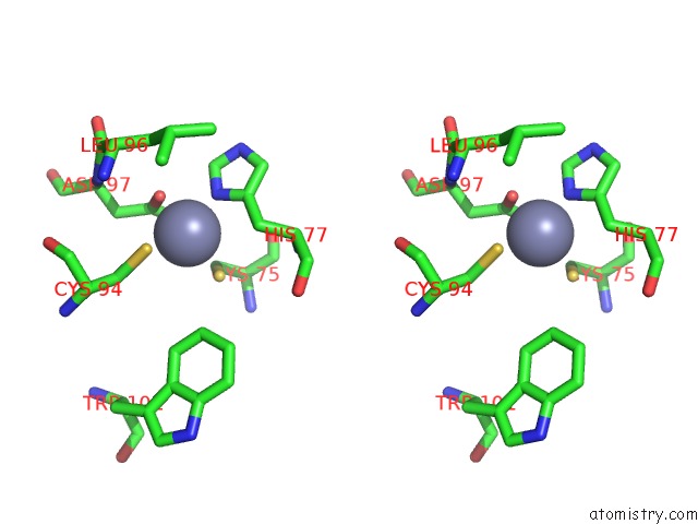
Stereo pair view
|
A full contact list of Zinc with other atoms in the Zn binding
site number 5 of Structure of A Glomulin-RBX1-CUL1 Complex within 5.0Å range:
probe
|
atom
|
residue
|
distance (Å)
|
B
|
Occ
|
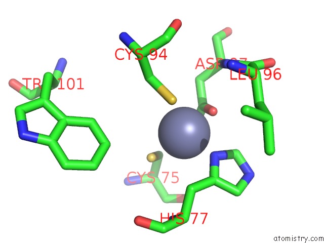
D:Zn202
b:0.3
occ:1.00
|
OD1
|
D:ASP97
|
1.9
|
0.1
|
1.0
|
CG
|
D:ASP97
|
2.0
|
0.8
|
1.0
|
OD2
|
D:ASP97
|
2.0
|
0.7
|
1.0
|
ND1
|
D:HIS77
|
2.1
|
0.7
|
1.0
|
SG
|
D:CYS75
|
2.3
|
0.9
|
1.0
|
SG
|
D:CYS94
|
2.5
|
0.7
|
1.0
|
CE1
|
D:HIS77
|
3.0
|
0.3
|
1.0
|
CB
|
D:CYS75
|
3.0
|
0.1
|
1.0
|
CG
|
D:HIS77
|
3.1
|
0.3
|
1.0
|
CB
|
D:ASP97
|
3.3
|
0.2
|
1.0
|
CB
|
D:HIS77
|
3.5
|
0.2
|
1.0
|
CB
|
D:CYS94
|
3.6
|
86.2
|
1.0
|
NE2
|
D:HIS77
|
4.1
|
1.0
|
1.0
|
CD2
|
D:HIS77
|
4.2
|
0.5
|
1.0
|
N
|
D:ASP97
|
4.3
|
0.2
|
1.0
|
CA
|
D:CYS75
|
4.3
|
0.2
|
1.0
|
O
|
D:CYS75
|
4.4
|
0.0
|
1.0
|
CA
|
D:ASP97
|
4.4
|
0.2
|
1.0
|
CZ3
|
D:TRP101
|
4.5
|
0.9
|
1.0
|
C
|
D:CYS75
|
4.5
|
0.5
|
1.0
|
N
|
D:HIS77
|
4.6
|
0.6
|
1.0
|
CA
|
D:HIS77
|
4.7
|
0.9
|
1.0
|
CB
|
D:LEU96
|
4.7
|
0.3
|
1.0
|
CA
|
D:CYS94
|
5.0
|
85.2
|
1.0
|
|
Zinc binding site 6 out
of 6 in 4f52
Go back to
Zinc Binding Sites List in 4f52
Zinc binding site 6 out
of 6 in the Structure of A Glomulin-RBX1-CUL1 Complex
Mono view
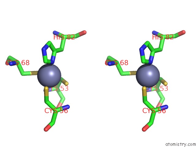
Stereo pair view
|
A full contact list of Zinc with other atoms in the Zn binding
site number 6 of Structure of A Glomulin-RBX1-CUL1 Complex within 5.0Å range:
probe
|
atom
|
residue
|
distance (Å)
|
B
|
Occ
|
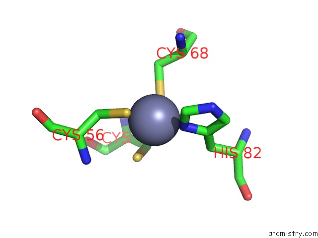
D:Zn203
b:0.7
occ:1.00
|
ND1
|
D:HIS82
|
1.9
|
0.7
|
1.0
|
SG
|
D:CYS68
|
2.0
|
97.6
|
1.0
|
SG
|
D:CYS56
|
2.3
|
90.5
|
1.0
|
SG
|
D:CYS53
|
2.5
|
0.2
|
1.0
|
CE1
|
D:HIS82
|
2.7
|
0.9
|
1.0
|
CG
|
D:HIS82
|
2.9
|
0.5
|
1.0
|
CB
|
D:CYS56
|
3.0
|
94.0
|
1.0
|
CB
|
D:CYS53
|
3.0
|
93.8
|
1.0
|
CB
|
D:CYS68
|
3.4
|
0.9
|
1.0
|
CB
|
D:HIS82
|
3.4
|
0.9
|
1.0
|
N
|
D:CYS53
|
3.8
|
94.4
|
1.0
|
NE2
|
D:HIS82
|
3.9
|
0.7
|
1.0
|
CD2
|
D:HIS82
|
4.0
|
0.8
|
1.0
|
CA
|
D:CYS53
|
4.0
|
92.5
|
1.0
|
CA
|
D:CYS68
|
4.1
|
0.2
|
1.0
|
CA
|
D:CYS56
|
4.2
|
99.7
|
1.0
|
N
|
D:CYS56
|
4.2
|
99.1
|
1.0
|
N
|
D:CYS68
|
4.6
|
0.9
|
1.0
|
C
|
D:CYS53
|
4.7
|
92.8
|
1.0
|
CA
|
D:HIS82
|
4.9
|
0.8
|
1.0
|
O
|
D:CYS53
|
5.0
|
0.8
|
1.0
|
|
Reference:
D.M.Duda,
J.L.Olszewski,
A.E.Tron,
M.Hammel,
L.J.Lambert,
M.B.Waddell,
T.Mittag,
J.A.Decaprio,
B.A.Schulman.
Structure of A Glomulin-RBX1-CUL1 Complex: Inhibition of A Ring E3 Ligase Through Masking of Its E2-Binding Surface. Mol.Cell V. 47 371 2012.
ISSN: ISSN 1097-2765
PubMed: 22748924
DOI: 10.1016/J.MOLCEL.2012.05.044
Page generated: Sat Oct 26 22:16:57 2024
|