Atomistry » Zinc » PDB 4cis-4csp » 4co9
Atomistry »
  Zinc »
    PDB 4cis-4csp »
      4co9 »

Zinc in PDB 4co9: Crystal Structure of Kynurenine Formamidase From Bacillus Anthracis

Enzymatic activity of Crystal Structure of Kynurenine Formamidase From Bacillus Anthracis

All present enzymatic activity of Crystal Structure of Kynurenine Formamidase From Bacillus Anthracis:
3.5.1.9;

Protein crystallography data

The structure of Crystal Structure of Kynurenine Formamidase From Bacillus Anthracis, PDB code: 4co9 was solved by L.Diaz-Saez, V.Srikannathasan, M.Zoltner, W.N.Hunter, with X-Ray Crystallography technique. A brief refinement statistics is given in the table below:

Resolution Low / High (Å) 83.76 / 1.95
Space group P 1 21 1
Cell size a, b, c (Å), α, β, γ (°) 73.710, 66.020, 83.760, 90.00, 90.32, 90.00
R / Rfree (%) 17.145 / 20.663

Other elements in 4co9:

The structure of Crystal Structure of Kynurenine Formamidase From Bacillus Anthracis also contains other interesting chemical elements:

Magnesium (Mg) 5 atoms

Zinc Binding Sites:

The binding sites of Zinc atom in the Crystal Structure of Kynurenine Formamidase From Bacillus Anthracis (pdb code 4co9). This binding sites where shown within 5.0 Angstroms radius around Zinc atom.
In total 8 binding sites of Zinc where determined in the Crystal Structure of Kynurenine Formamidase From Bacillus Anthracis, PDB code: 4co9:
Jump to Zinc binding site number: 1; 2; 3; 4; 5; 6; 7; 8;

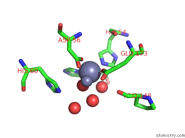
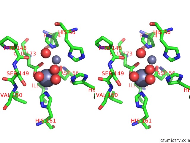
Zinc binding site 1 out of 8 in 4co9

Go back to Zinc Binding Sites List in 4co9
Zinc binding site 1 out of 8 in the Crystal Structure of Kynurenine Formamidase From Bacillus Anthracis


Mono view


Stereo pair view

A full contact list of Zinc with other atoms in the Zn binding site number 1 of Crystal Structure of Kynurenine Formamidase From Bacillus Anthracis within 5.0Å range:
probe atom residue distance (Å) B Occ
A:Zn401

b:19.9
occ:1.00
OD2 A:ASP56 2.0 9.1 1.0
O A:HOH2108 2.0 10.2 1.0
OE2 A:GLU173 2.1 7.0 1.0
NE2 A:HIS161 2.3 10.2 1.0
O A:HOH2238 2.5 22.6 1.0
OE1 A:GLU173 2.6 6.3 1.0
CD A:GLU173 2.6 6.5 1.0
CG A:ASP56 3.0 8.9 1.0
ZN A:ZN402 3.1 23.9 1.0
CE1 A:HIS161 3.2 10.1 1.0
CD2 A:HIS161 3.3 10.4 1.0
OD1 A:ASP56 3.3 9.6 1.0
O A:HOH2044 4.0 7.3 1.0
CG A:GLU173 4.1 6.3 1.0
O A:HOH2112 4.1 23.0 1.0
CB A:ASP56 4.3 8.8 1.0
CD2 A:HIS60 4.3 10.0 1.0
ND1 A:HIS161 4.3 10.5 1.0
O A:PRO148 4.3 8.9 1.0
CG A:HIS161 4.4 10.6 1.0
OG A:SER149 4.4 9.7 1.0
NE2 A:HIS60 4.4 10.0 1.0
CG2 A:ILE171 4.6 8.2 1.0
CA A:SER149 4.6 9.5 1.0
N A:VAL150 4.9 10.0 1.0
ND1 A:HIS50 4.9 8.5 1.0
CB A:SER149 4.9 9.8 1.0
NE2 A:HIS54 5.0 7.4 1.0

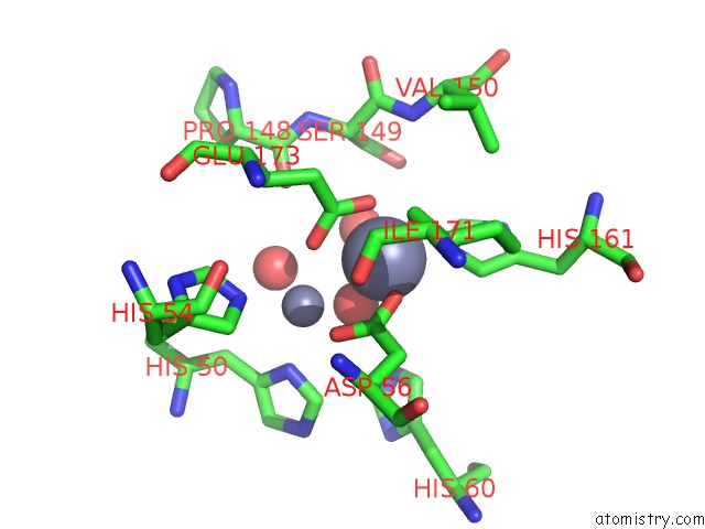
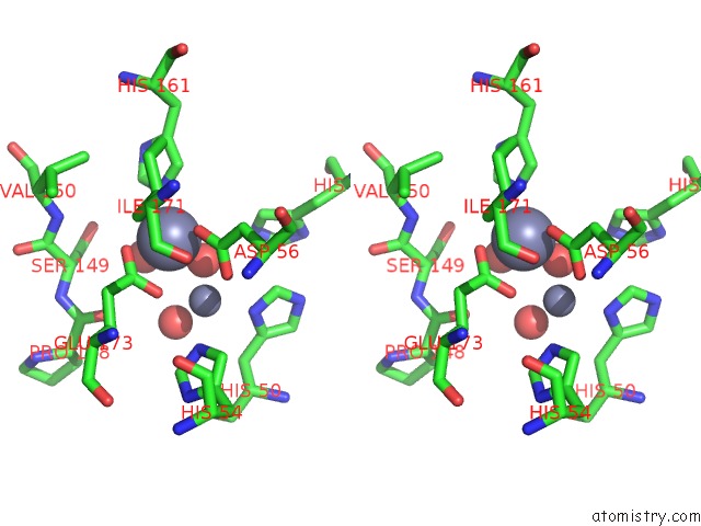
Zinc binding site 2 out of 8 in 4co9

Go back to Zinc Binding Sites List in 4co9
Zinc binding site 2 out of 8 in the Crystal Structure of Kynurenine Formamidase From Bacillus Anthracis


Mono view


Stereo pair view

A full contact list of Zinc with other atoms in the Zn binding site number 2 of Crystal Structure of Kynurenine Formamidase From Bacillus Anthracis within 5.0Å range:
probe atom residue distance (Å) B Occ
A:Zn402

b:23.9
occ:1.00
O A:HOH2108 2.0 10.2 1.0
OD1 A:ASP56 2.1 9.6 1.0
ND1 A:HIS50 2.2 8.5 1.0
NE2 A:HIS54 2.3 7.4 1.0
OE1 A:GLU173 2.4 6.3 1.0
O A:HOH2044 2.5 7.3 1.0
CG A:ASP56 3.0 8.9 1.0
CE1 A:HIS50 3.1 8.6 1.0
ZN A:ZN401 3.1 19.9 1.0
CD2 A:HIS54 3.1 7.2 1.0
OD2 A:ASP56 3.3 9.1 1.0
CG A:HIS50 3.3 8.5 1.0
CE1 A:HIS54 3.4 7.3 1.0
CD A:GLU173 3.6 6.5 1.0
CB A:HIS50 3.7 8.7 1.0
O A:HOH2238 4.1 22.6 1.0
OE2 A:GLU173 4.1 7.0 1.0
NE2 A:HIS50 4.2 8.7 1.0
CG A:HIS54 4.3 7.2 1.0
CD2 A:HIS50 4.4 8.7 1.0
ND1 A:HIS54 4.4 7.3 1.0
CB A:ASP56 4.4 8.8 1.0
NE2 A:HIS60 4.5 10.0 1.0
O A:PRO148 4.5 8.9 1.0
CA A:HIS50 4.5 8.7 1.0
CD2 A:HIS60 4.6 10.0 1.0
CG A:GLU173 4.7 6.3 1.0
CB A:GLU173 4.9 6.2 1.0
O A:HOH2112 4.9 23.0 1.0
CA A:ASP56 4.9 8.5 1.0

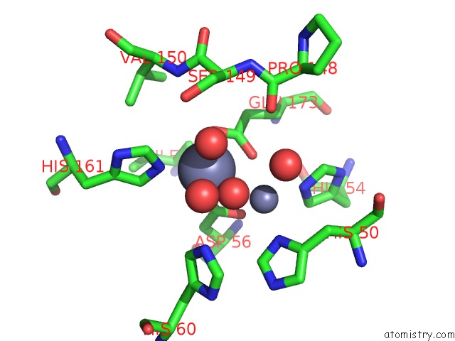
Zinc binding site 3 out of 8 in 4co9

Go back to Zinc Binding Sites List in 4co9
Zinc binding site 3 out of 8 in the Crystal Structure of Kynurenine Formamidase From Bacillus Anthracis


Mono view


Stereo pair view

A full contact list of Zinc with other atoms in the Zn binding site number 3 of Crystal Structure of Kynurenine Formamidase From Bacillus Anthracis within 5.0Å range:
probe atom residue distance (Å) B Occ
B:Zn401

b:21.1
occ:1.00
OD2 B:ASP56 2.0 9.6 1.0
O B:HOH2073 2.1 9.9 1.0
OE2 B:GLU173 2.1 8.7 1.0
NE2 B:HIS161 2.2 13.8 1.0
O B:HOH2147 2.5 20.5 1.0
OE1 B:GLU173 2.6 8.3 1.0
CD B:GLU173 2.7 8.8 1.0
CG B:ASP56 3.0 9.5 1.0
ZN B:ZN402 3.1 22.1 1.0
CE1 B:HIS161 3.1 14.2 1.0
CD2 B:HIS161 3.2 13.6 1.0
OD1 B:ASP56 3.4 9.1 1.0
O B:HOH2037 3.9 11.4 1.0
CG B:GLU173 4.1 8.5 1.0
ND1 B:HIS161 4.3 13.8 1.0
O B:PRO148 4.3 14.5 1.0
CG B:HIS161 4.3 14.2 1.0
CB B:ASP56 4.4 9.5 1.0
OG B:SER149 4.4 14.5 1.0
CD2 B:HIS60 4.5 17.6 1.0
CG2 B:ILE171 4.6 10.1 1.0
CA B:SER149 4.6 13.9 1.0
NE2 B:HIS60 4.7 17.8 1.0
N B:VAL150 4.9 15.2 1.0
ND1 B:HIS50 4.9 11.9 1.0
CB B:SER149 4.9 14.3 1.0
NE2 B:HIS54 5.0 8.3 1.0

Zinc binding site 4 out of 8 in 4co9

Go back to Zinc Binding Sites List in 4co9
Zinc binding site 4 out of 8 in the Crystal Structure of Kynurenine Formamidase From Bacillus Anthracis


Mono view


Stereo pair view

A full contact list of Zinc with other atoms in the Zn binding site number 4 of Crystal Structure of Kynurenine Formamidase From Bacillus Anthracis within 5.0Å range:
probe atom residue distance (Å) B Occ
B:Zn402

b:22.1
occ:1.00
O B:HOH2073 2.1 9.9 1.0
OD1 B:ASP56 2.2 9.1 1.0
NE2 B:HIS54 2.3 8.3 1.0
OE1 B:GLU173 2.3 8.3 1.0
ND1 B:HIS50 2.3 11.9 1.0
O B:HOH2037 2.3 11.4 1.0
ZN B:ZN401 3.1 21.1 1.0
CG B:ASP56 3.1 9.5 1.0
CD2 B:HIS54 3.1 8.1 1.0
CE1 B:HIS50 3.2 12.4 1.0
OD2 B:ASP56 3.3 9.6 1.0
CE1 B:HIS54 3.3 8.1 1.0
CG B:HIS50 3.4 12.6 1.0
CD B:GLU173 3.5 8.8 1.0
CB B:HIS50 3.7 12.8 1.0
O B:HOH2147 4.0 20.5 1.0
OE2 B:GLU173 4.0 8.7 1.0
CG B:HIS54 4.3 8.1 1.0
NE2 B:HIS50 4.3 12.4 1.0
ND1 B:HIS54 4.4 7.9 1.0
O B:PRO148 4.4 14.5 1.0
CD2 B:HIS50 4.4 12.4 1.0
CB B:ASP56 4.5 9.5 1.0
CA B:HIS50 4.5 13.2 1.0
CG B:GLU173 4.7 8.5 1.0
CB B:GLU173 4.8 8.4 1.0
NE2 B:HIS60 4.8 17.8 1.0
CD2 B:HIS60 4.9 17.6 1.0
CA B:ASP56 4.9 9.7 1.0

Zinc binding site 5 out of 8 in 4co9

Go back to Zinc Binding Sites List in 4co9
Zinc binding site 5 out of 8 in the Crystal Structure of Kynurenine Formamidase From Bacillus Anthracis


Mono view


Stereo pair view

A full contact list of Zinc with other atoms in the Zn binding site number 5 of Crystal Structure of Kynurenine Formamidase From Bacillus Anthracis within 5.0Å range:
probe atom residue distance (Å) B Occ
C:Zn401

b:23.3
occ:1.00
OD2 C:ASP56 2.0 9.5 1.0
O C:HOH2102 2.0 9.6 1.0
OE2 C:GLU173 2.1 8.7 1.0
NE2 C:HIS161 2.3 12.6 1.0
O C:HOH2201 2.5 18.8 1.0
OE1 C:GLU173 2.6 8.4 1.0
CD C:GLU173 2.7 8.7 1.0
CG C:ASP56 3.0 8.7 1.0
ZN C:ZN402 3.1 22.1 1.0
CE1 C:HIS161 3.2 12.8 1.0
CD2 C:HIS161 3.3 12.9 1.0
OD1 C:ASP56 3.3 8.4 1.0
O C:HOH2105 3.9 22.9 1.0
O C:HOH2048 4.0 10.4 1.0
CG C:GLU173 4.2 8.5 1.0
CD2 C:HIS60 4.3 10.8 1.0
CB C:ASP56 4.3 8.5 1.0
O C:PRO148 4.3 12.3 1.0
ND1 C:HIS161 4.3 13.0 1.0
OG C:SER149 4.4 12.7 1.0
NE2 C:HIS60 4.4 10.8 1.0
CG C:HIS161 4.4 13.1 1.0
CG2 C:ILE171 4.6 10.4 1.0
CA C:SER149 4.6 12.5 1.0
ND1 C:HIS50 4.8 8.6 1.0
N C:VAL150 4.9 13.0 1.0
CB C:SER149 4.9 12.7 1.0
NE2 C:HIS54 5.0 7.5 1.0

Zinc binding site 6 out of 8 in 4co9

Go back to Zinc Binding Sites List in 4co9
Zinc binding site 6 out of 8 in the Crystal Structure of Kynurenine Formamidase From Bacillus Anthracis


Mono view


Stereo pair view

A full contact list of Zinc with other atoms in the Zn binding site number 6 of Crystal Structure of Kynurenine Formamidase From Bacillus Anthracis within 5.0Å range:
probe atom residue distance (Å) B Occ
C:Zn402

b:22.1
occ:1.00
O C:HOH2102 2.1 9.6 1.0
OD1 C:ASP56 2.1 8.4 1.0
ND1 C:HIS50 2.2 8.6 1.0
NE2 C:HIS54 2.3 7.5 1.0
OE1 C:GLU173 2.4 8.4 1.0
O C:HOH2048 2.6 10.4 1.0
CE1 C:HIS50 3.0 8.6 1.0
CG C:ASP56 3.0 8.7 1.0
CD2 C:HIS54 3.1 7.3 1.0
ZN C:ZN401 3.1 23.3 1.0
OD2 C:ASP56 3.3 9.5 1.0
CG C:HIS50 3.3 8.5 1.0
CE1 C:HIS54 3.4 7.5 1.0
CD C:GLU173 3.6 8.7 1.0
CB C:HIS50 3.7 8.6 1.0
OE2 C:GLU173 4.1 8.7 1.0
NE2 C:HIS50 4.2 8.6 1.0
CG C:HIS54 4.3 7.3 1.0
CD2 C:HIS50 4.4 8.4 1.0
O C:HOH2201 4.4 18.8 1.0
CB C:ASP56 4.4 8.5 1.0
ND1 C:HIS54 4.4 7.4 1.0
NE2 C:HIS60 4.5 10.8 1.0
CA C:HIS50 4.5 8.5 1.0
O C:PRO148 4.5 12.3 1.0
CD2 C:HIS60 4.6 10.8 1.0
CG C:GLU173 4.8 8.5 1.0
CA C:ASP56 4.8 8.4 1.0
CB C:GLU173 4.9 8.4 1.0
O C:HOH2105 4.9 22.9 1.0
N C:ASP56 5.0 8.0 1.0

Zinc binding site 7 out of 8 in 4co9

Go back to Zinc Binding Sites List in 4co9
Zinc binding site 7 out of 8 in the Crystal Structure of Kynurenine Formamidase From Bacillus Anthracis


Mono view


Stereo pair view

A full contact list of Zinc with other atoms in the Zn binding site number 7 of Crystal Structure of Kynurenine Formamidase From Bacillus Anthracis within 5.0Å range:
probe atom residue distance (Å) B Occ
D:Zn401

b:17.5
occ:1.00
O D:HOH2075 2.0 10.8 1.0
OD2 D:ASP56 2.0 7.5 1.0
OE2 D:GLU173 2.1 7.8 1.0
NE2 D:HIS161 2.2 9.8 1.0
O D:HOH2163 2.4 18.2 1.0
OE1 D:GLU173 2.6 7.4 1.0
CD D:GLU173 2.7 7.7 1.0
CG D:ASP56 3.0 7.5 1.0
ZN D:ZN402 3.1 21.9 1.0
CE1 D:HIS161 3.1 9.8 1.0
CD2 D:HIS161 3.2 10.0 1.0
OD1 D:ASP56 3.4 7.4 1.0
O D:HOH2036 4.0 11.1 1.0
O D:HOH2039 4.0 40.5 1.0
CG D:GLU173 4.1 7.5 1.0
ND1 D:HIS161 4.3 9.8 1.0
CB D:ASP56 4.3 7.4 1.0
CG D:HIS161 4.3 9.9 1.0
O D:PRO148 4.3 11.4 1.0
OG D:SER149 4.4 9.7 1.0
CD2 D:HIS60 4.5 10.3 1.0
CG2 D:ILE171 4.6 8.5 1.0
CA D:SER149 4.6 10.0 1.0
NE2 D:HIS60 4.7 10.5 1.0
ND1 D:HIS50 4.9 11.3 1.0
N D:VAL150 4.9 10.6 1.0
CB D:SER149 4.9 9.9 1.0
NE2 D:HIS54 5.0 6.7 1.0

Zinc binding site 8 out of 8 in 4co9

Go back to Zinc Binding Sites List in 4co9
Zinc binding site 8 out of 8 in the Crystal Structure of Kynurenine Formamidase From Bacillus Anthracis


Mono view


Stereo pair view

A full contact list of Zinc with other atoms in the Zn binding site number 8 of Crystal Structure of Kynurenine Formamidase From Bacillus Anthracis within 5.0Å range:
probe atom residue distance (Å) B Occ
D:Zn402

b:21.9
occ:1.00
O D:HOH2075 2.1 10.8 1.0
OD1 D:ASP56 2.2 7.4 1.0
OE1 D:GLU173 2.3 7.4 1.0
NE2 D:HIS54 2.3 6.7 1.0
ND1 D:HIS50 2.3 11.3 1.0
O D:HOH2036 2.4 11.1 1.0
ZN D:ZN401 3.1 17.5 1.0
CG D:ASP56 3.1 7.5 1.0
CD2 D:HIS54 3.1 6.5 1.0
CE1 D:HIS50 3.2 11.4 1.0
OD2 D:ASP56 3.3 7.5 1.0
CE1 D:HIS54 3.3 6.7 1.0
CG D:HIS50 3.4 11.4 1.0
CD D:GLU173 3.4 7.7 1.0
O D:HOH2039 3.7 40.5 1.0
CB D:HIS50 3.7 11.5 1.0
OE2 D:GLU173 4.0 7.8 1.0
O D:HOH2163 4.1 18.2 1.0
CG D:HIS54 4.3 6.5 1.0
O D:PRO148 4.4 11.4 1.0
NE2 D:HIS50 4.4 11.7 1.0
ND1 D:HIS54 4.4 6.5 1.0
CB D:ASP56 4.5 7.4 1.0
CD2 D:HIS50 4.5 11.4 1.0
CA D:HIS50 4.5 11.3 1.0
CG D:GLU173 4.6 7.5 1.0
CB D:GLU173 4.7 7.3 1.0
NE2 D:HIS60 4.8 10.5 1.0
CD2 D:HIS60 4.8 10.3 1.0
O D:HOH2038 4.9 28.2 1.0
CA D:ASP56 4.9 7.5 1.0

Reference:

L.Diaz-Saez, V.Srikannathasan, M.Zoltner, W.N.Hunter. Structure of Bacterial Kynurenine Formamidase Reveals A Crowded Binuclear-Zinc Catalytic Site Primed to Generate A Potent Nucleophile. Biochem.J. V. 462 581 2014.
ISSN: ISSN 0264-6021
PubMed: 24942958
DOI: 10.1042/BJ20140511
Page generated: Sat Oct 26 20:53:23 2024

Last articles

Zn in 9J0N
Zn in 9J0O
Zn in 9J0P
Zn in 9FJX
Zn in 9EKB
Zn in 9C0F
Zn in 9CAH
Zn in 9CH0
Zn in 9CH3
Zn in 9CH1
© Copyright 2008-2020 by atomistry.com
Home   |    Site Map   |    Copyright   |    Contact us   |    Privacy