Zinc »
PDB 4bjb-4bud »
4bpu »
Zinc in PDB 4bpu: Crystal Structure of Human Primase in Heterodimeric Form, Comprising Pris and Truncated Pril Lacking the C-Terminal Fe-S Domain.
Protein crystallography data
The structure of Crystal Structure of Human Primase in Heterodimeric Form, Comprising Pris and Truncated Pril Lacking the C-Terminal Fe-S Domain., PDB code: 4bpu
was solved by
M.L.Kilkenny,
R.L.Perera,
L.Pellegrini,
with X-Ray Crystallography technique. A brief refinement statistics is given in the table below:
| Resolution Low / High (Å) | 29.617 / 2.70 |
| Space group | P 1 21 1 |
| Cell size a, b, c (Å), α, β, γ (°) | 114.550, 68.640, 126.760, 90.00, 104.36, 90.00 |
| R / Rfree (%) | 20.28 / 24.54 |
Zinc Binding Sites:
The binding sites of Zinc atom in the Crystal Structure of Human Primase in Heterodimeric Form, Comprising Pris and Truncated Pril Lacking the C-Terminal Fe-S Domain.
(pdb code 4bpu). This binding sites where shown within
5.0 Angstroms radius around Zinc atom.
In total 2 binding sites of Zinc where determined in the Crystal Structure of Human Primase in Heterodimeric Form, Comprising Pris and Truncated Pril Lacking the C-Terminal Fe-S Domain., PDB code: 4bpu:
Jump to Zinc binding site number: 1; 2;
In total 2 binding sites of Zinc where determined in the Crystal Structure of Human Primase in Heterodimeric Form, Comprising Pris and Truncated Pril Lacking the C-Terminal Fe-S Domain., PDB code: 4bpu:
Jump to Zinc binding site number: 1; 2;
Zinc binding site 1 out of 2 in 4bpu
Go back to
Zinc binding site 1 out
of 2 in the Crystal Structure of Human Primase in Heterodimeric Form, Comprising Pris and Truncated Pril Lacking the C-Terminal Fe-S Domain.
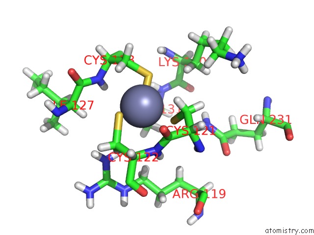
Mono view
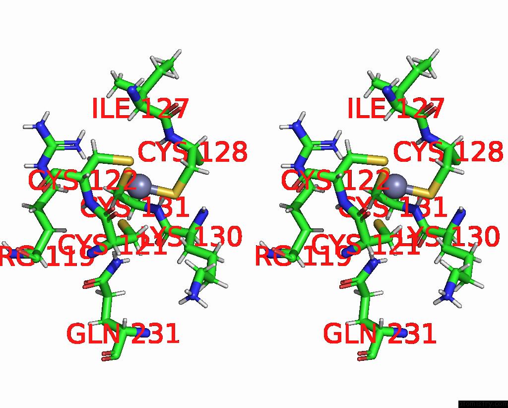
Stereo pair view
Mono view
Stereo pair view
A full contact list of Zinc with other atoms in the Zn binding
site number 1 of Crystal Structure of Human Primase in Heterodimeric Form, Comprising Pris and Truncated Pril Lacking the C-Terminal Fe-S Domain. within 5.0Å range:
|
Zinc binding site 2 out of 2 in 4bpu
Go back to
Zinc binding site 2 out
of 2 in the Crystal Structure of Human Primase in Heterodimeric Form, Comprising Pris and Truncated Pril Lacking the C-Terminal Fe-S Domain.
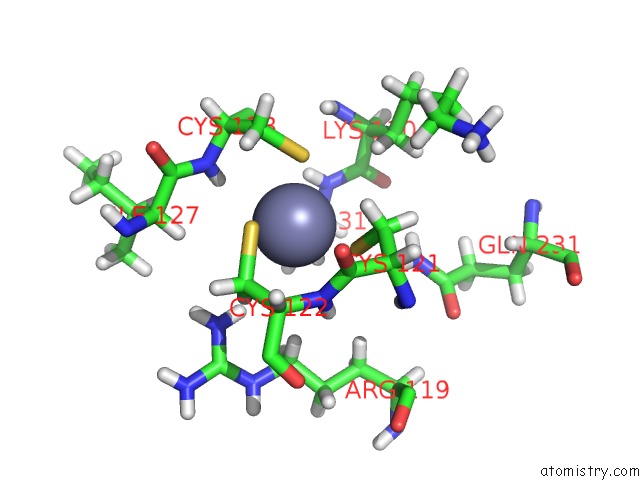
Mono view
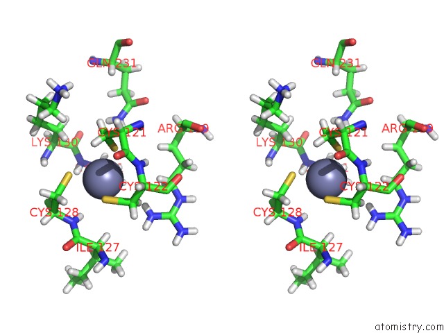
Stereo pair view
Mono view
Stereo pair view
A full contact list of Zinc with other atoms in the Zn binding
site number 2 of Crystal Structure of Human Primase in Heterodimeric Form, Comprising Pris and Truncated Pril Lacking the C-Terminal Fe-S Domain. within 5.0Å range:
|
Reference:
M.L.Kilkenny,
M.Longo,
R.L.Perera,
L.Pellegrini.
Structures of Human Primase Reveal Design of Nucleotide Elongation Site and Mode of Pol Alpha Tethering Proc.Natl.Acad.Sci.Usa V. 110 15961 2013.
ISSN: ISSN 0027-8424
PubMed: 24043831
DOI: 10.1073/PNAS.1311185110
Page generated: Sat Oct 26 19:54:22 2024
ISSN: ISSN 0027-8424
PubMed: 24043831
DOI: 10.1073/PNAS.1311185110
Last articles
Zn in 9MJ5Zn in 9HNW
Zn in 9G0L
Zn in 9FNE
Zn in 9DZN
Zn in 9E0I
Zn in 9D32
Zn in 9DAK
Zn in 8ZXC
Zn in 8ZUF