Zinc »
PDB 4bjb-4bud »
4bp0 »
Zinc in PDB 4bp0: Crystal Structure of the Closed Form of Pseudomonas Aeruginosa Spm-1
Enzymatic activity of Crystal Structure of the Closed Form of Pseudomonas Aeruginosa Spm-1
All present enzymatic activity of Crystal Structure of the Closed Form of Pseudomonas Aeruginosa Spm-1:
3.5.2.6;
3.5.2.6;
Protein crystallography data
The structure of Crystal Structure of the Closed Form of Pseudomonas Aeruginosa Spm-1, PDB code: 4bp0
was solved by
M.A.Mcdonough,
J.Brem,
C.J.Schofield,
with X-Ray Crystallography technique. A brief refinement statistics is given in the table below:
| Resolution Low / High (Å) | 71.9428 / 2.24 |
| Space group | P 21 21 21 |
| Cell size a, b, c (Å), α, β, γ (°) | 45.650, 83.550, 282.410, 90.00, 90.00, 90.00 |
| R / Rfree (%) | 15.16 / 19.81 |
Other elements in 4bp0:
The structure of Crystal Structure of the Closed Form of Pseudomonas Aeruginosa Spm-1 also contains other interesting chemical elements:
| Chlorine | (Cl) | 7 atoms |
Zinc Binding Sites:
The binding sites of Zinc atom in the Crystal Structure of the Closed Form of Pseudomonas Aeruginosa Spm-1
(pdb code 4bp0). This binding sites where shown within
5.0 Angstroms radius around Zinc atom.
In total 10 binding sites of Zinc where determined in the Crystal Structure of the Closed Form of Pseudomonas Aeruginosa Spm-1, PDB code: 4bp0:
Jump to Zinc binding site number: 1; 2; 3; 4; 5; 6; 7; 8; 9; 10;
In total 10 binding sites of Zinc where determined in the Crystal Structure of the Closed Form of Pseudomonas Aeruginosa Spm-1, PDB code: 4bp0:
Jump to Zinc binding site number: 1; 2; 3; 4; 5; 6; 7; 8; 9; 10;
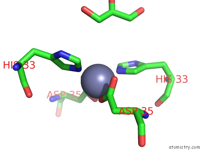
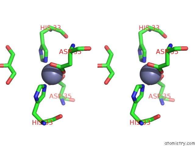
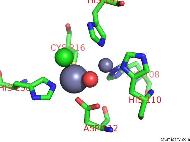
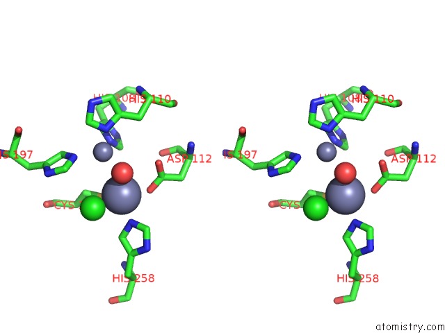
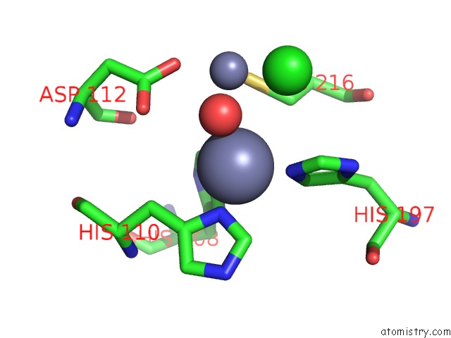
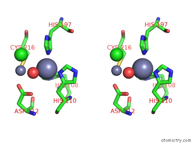
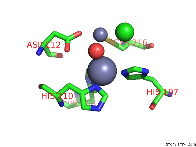
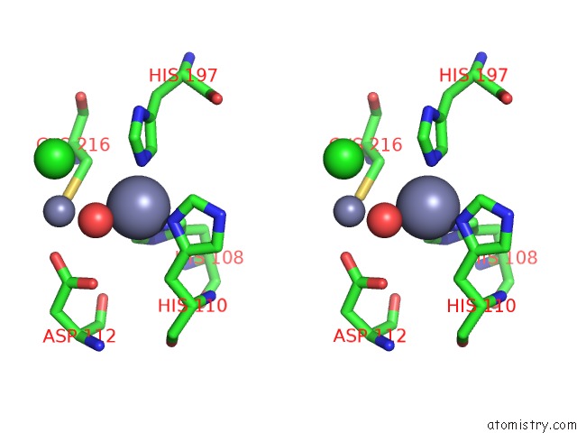
Zinc binding site 1 out of 10 in 4bp0
Go back to
Zinc binding site 1 out
of 10 in the Crystal Structure of the Closed Form of Pseudomonas Aeruginosa Spm-1
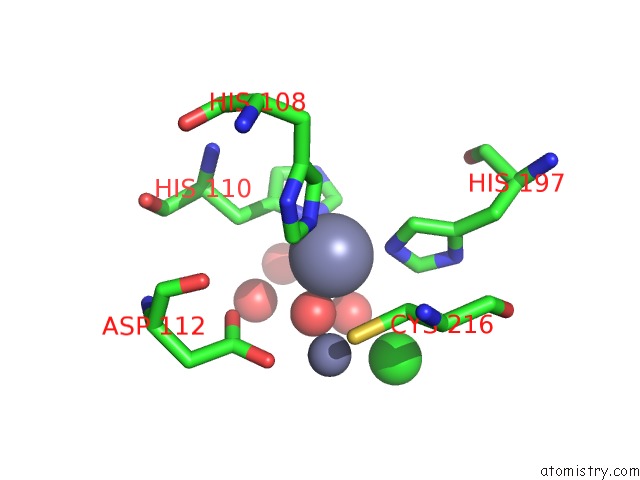
Mono view
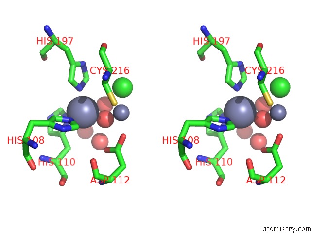
Stereo pair view
Mono view
Stereo pair view
A full contact list of Zinc with other atoms in the Zn binding
site number 1 of Crystal Structure of the Closed Form of Pseudomonas Aeruginosa Spm-1 within 5.0Å range:
|
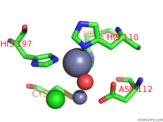
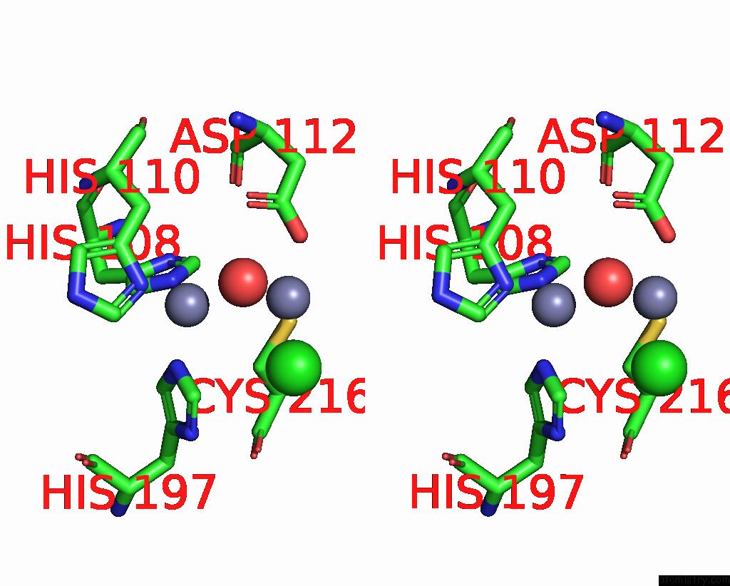
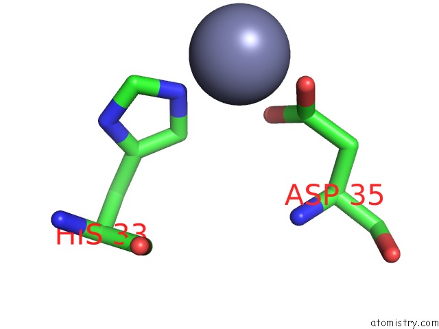
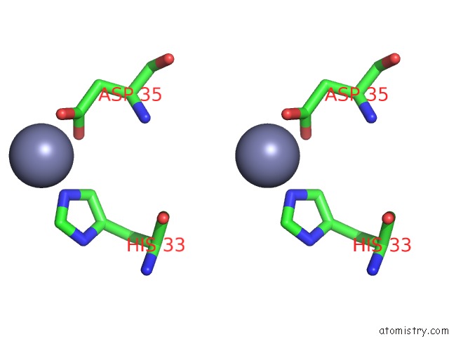
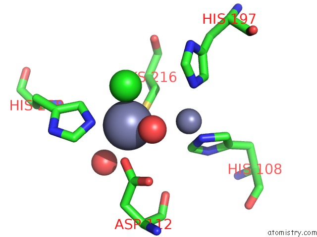
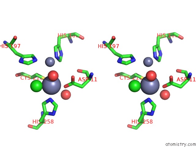
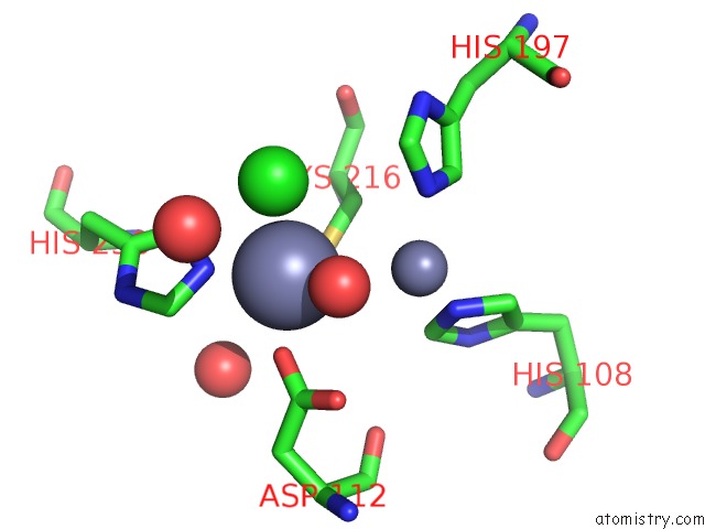
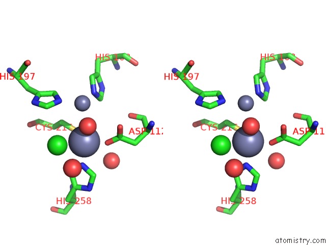
Zinc binding site 2 out of 10 in 4bp0
Go back to
Zinc binding site 2 out
of 10 in the Crystal Structure of the Closed Form of Pseudomonas Aeruginosa Spm-1
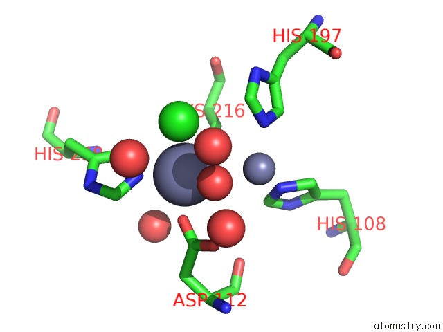
Mono view
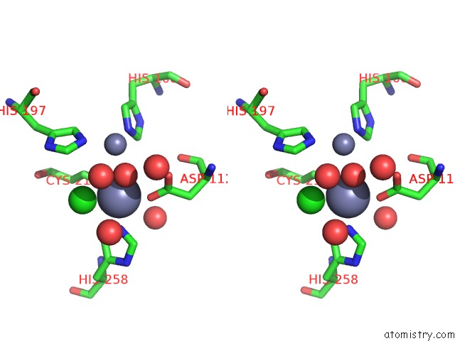
Stereo pair view
Mono view
Stereo pair view
A full contact list of Zinc with other atoms in the Zn binding
site number 2 of Crystal Structure of the Closed Form of Pseudomonas Aeruginosa Spm-1 within 5.0Å range:
|
Zinc binding site 3 out of 10 in 4bp0
Go back to
Zinc binding site 3 out
of 10 in the Crystal Structure of the Closed Form of Pseudomonas Aeruginosa Spm-1
Mono view
Stereo pair view
Mono view
Stereo pair view
A full contact list of Zinc with other atoms in the Zn binding
site number 3 of Crystal Structure of the Closed Form of Pseudomonas Aeruginosa Spm-1 within 5.0Å range:
|
Zinc binding site 4 out of 10 in 4bp0
Go back to
Zinc binding site 4 out
of 10 in the Crystal Structure of the Closed Form of Pseudomonas Aeruginosa Spm-1
Mono view
Stereo pair view
Mono view
Stereo pair view
A full contact list of Zinc with other atoms in the Zn binding
site number 4 of Crystal Structure of the Closed Form of Pseudomonas Aeruginosa Spm-1 within 5.0Å range:
|
Zinc binding site 5 out of 10 in 4bp0
Go back to
Zinc binding site 5 out
of 10 in the Crystal Structure of the Closed Form of Pseudomonas Aeruginosa Spm-1
Mono view
Stereo pair view
Mono view
Stereo pair view
A full contact list of Zinc with other atoms in the Zn binding
site number 5 of Crystal Structure of the Closed Form of Pseudomonas Aeruginosa Spm-1 within 5.0Å range:
|
Zinc binding site 6 out of 10 in 4bp0
Go back to
Zinc binding site 6 out
of 10 in the Crystal Structure of the Closed Form of Pseudomonas Aeruginosa Spm-1
Mono view
Stereo pair view
Mono view
Stereo pair view
A full contact list of Zinc with other atoms in the Zn binding
site number 6 of Crystal Structure of the Closed Form of Pseudomonas Aeruginosa Spm-1 within 5.0Å range:
|
Zinc binding site 7 out of 10 in 4bp0
Go back to
Zinc binding site 7 out
of 10 in the Crystal Structure of the Closed Form of Pseudomonas Aeruginosa Spm-1
Mono view
Stereo pair view
Mono view
Stereo pair view
A full contact list of Zinc with other atoms in the Zn binding
site number 7 of Crystal Structure of the Closed Form of Pseudomonas Aeruginosa Spm-1 within 5.0Å range:
|
Zinc binding site 8 out of 10 in 4bp0
Go back to
Zinc binding site 8 out
of 10 in the Crystal Structure of the Closed Form of Pseudomonas Aeruginosa Spm-1
Mono view
Stereo pair view
Mono view
Stereo pair view
A full contact list of Zinc with other atoms in the Zn binding
site number 8 of Crystal Structure of the Closed Form of Pseudomonas Aeruginosa Spm-1 within 5.0Å range:
|
Zinc binding site 9 out of 10 in 4bp0
Go back to
Zinc binding site 9 out
of 10 in the Crystal Structure of the Closed Form of Pseudomonas Aeruginosa Spm-1
Mono view
Stereo pair view
Mono view
Stereo pair view
A full contact list of Zinc with other atoms in the Zn binding
site number 9 of Crystal Structure of the Closed Form of Pseudomonas Aeruginosa Spm-1 within 5.0Å range:
|
Zinc binding site 10 out of 10 in 4bp0
Go back to
Zinc binding site 10 out
of 10 in the Crystal Structure of the Closed Form of Pseudomonas Aeruginosa Spm-1
Mono view
Stereo pair view
Mono view
Stereo pair view
A full contact list of Zinc with other atoms in the Zn binding
site number 10 of Crystal Structure of the Closed Form of Pseudomonas Aeruginosa Spm-1 within 5.0Å range:
|
Reference:
M.A.Mcdonough,
J.Brem,
C.J.Schofield.
The Crystal Structure of the Closed Form or Pseudomonas Aeruginosa Spm-1 To Be Published.
Page generated: Sat Oct 26 19:52:31 2024
Last articles
Zn in 9MJ5Zn in 9HNW
Zn in 9G0L
Zn in 9FNE
Zn in 9DZN
Zn in 9E0I
Zn in 9D32
Zn in 9DAK
Zn in 8ZXC
Zn in 8ZUF