Zinc »
PDB 4ad9-4arf »
4aqy »
Zinc in PDB 4aqy: Structure of Ribosome-Apramycin Complexes
Protein crystallography data
The structure of Structure of Ribosome-Apramycin Complexes, PDB code: 4aqy
was solved by
T.Matt,
C.L.Ng,
K.Lang,
S.H.Sha,
R.Akbergenov,
D.Shcherbakov,
M.Meyer,
S.Duscha,
J.Xie,
S.R.Dubbaka,
D.Perez-Fernandez,
A.Vasella,
V.Ramakrishnan,
J.Schacht,
E.C.Bottger,
with X-Ray Crystallography technique. A brief refinement statistics is given in the table below:
| Resolution Low / High (Å) | 40.00 / 3.50 |
| Space group | P 41 21 2 |
| Cell size a, b, c (Å), α, β, γ (°) | 402.180, 402.180, 175.000, 90.00, 90.00, 90.00 |
| R / Rfree (%) | 19.3 / 23.5 |
Other elements in 4aqy:
The structure of Structure of Ribosome-Apramycin Complexes also contains other interesting chemical elements:
| Magnesium | (Mg) | 203 atoms |
| Potassium | (K) | 15 atoms |
Zinc Binding Sites:
The binding sites of Zinc atom in the Structure of Ribosome-Apramycin Complexes
(pdb code 4aqy). This binding sites where shown within
5.0 Angstroms radius around Zinc atom.
In total 2 binding sites of Zinc where determined in the Structure of Ribosome-Apramycin Complexes, PDB code: 4aqy:
Jump to Zinc binding site number: 1; 2;
In total 2 binding sites of Zinc where determined in the Structure of Ribosome-Apramycin Complexes, PDB code: 4aqy:
Jump to Zinc binding site number: 1; 2;
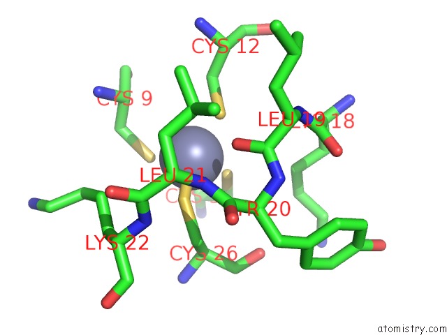
Zinc binding site 1 out of 2 in 4aqy
Go back to
Zinc binding site 1 out
of 2 in the Structure of Ribosome-Apramycin Complexes
Mono view

Stereo pair view
Mono view
Stereo pair view
A full contact list of Zinc with other atoms in the Zn binding
site number 1 of Structure of Ribosome-Apramycin Complexes within 5.0Å range:
|
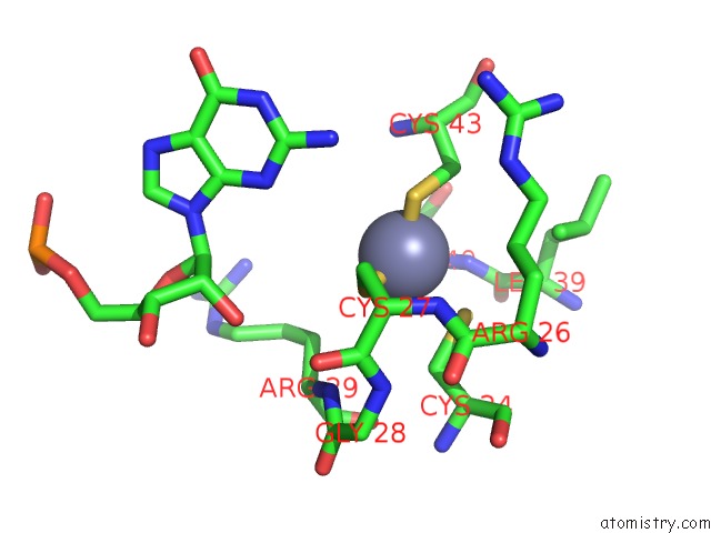
Zinc binding site 2 out of 2 in 4aqy
Go back to
Zinc binding site 2 out
of 2 in the Structure of Ribosome-Apramycin Complexes
Mono view

Stereo pair view
Mono view
Stereo pair view
A full contact list of Zinc with other atoms in the Zn binding
site number 2 of Structure of Ribosome-Apramycin Complexes within 5.0Å range:
|
Reference:
T.Matt,
C.L.Ng,
K.Lang,
S.H.Sha,
R.Akbergenov,
D.Shcherbakov,
M.Meyer,
S.Duscha,
J.Xie,
S.R.Dubbaka,
D.Perez-Fernandez,
A.Vasella,
V.Ramakrishnan,
J.Schacht,
E.C.Bottger.
Dissociation of Antibacterial Activity and Aminoglycoside Ototoxicity in the 4-Monosubstituted 2-Deoxystreptamine Apramycin. Proc.Natl.Acad.Sci.Usa V. 109 10984 2012.
ISSN: ISSN 0027-8424
PubMed: 22699498
DOI: 10.1073/PNAS.1204073109
Page generated: Sat Oct 26 19:21:09 2024
ISSN: ISSN 0027-8424
PubMed: 22699498
DOI: 10.1073/PNAS.1204073109
Last articles
Zn in 9MJ5Zn in 9HNW
Zn in 9G0L
Zn in 9FNE
Zn in 9DZN
Zn in 9E0I
Zn in 9D32
Zn in 9DAK
Zn in 8ZXC
Zn in 8ZUF