Zinc »
PDB 4ad9-4arf »
4aig »
Zinc in PDB 4aig: Adamalysin II with Phosphonate Inhibitor
Enzymatic activity of Adamalysin II with Phosphonate Inhibitor
All present enzymatic activity of Adamalysin II with Phosphonate Inhibitor:
3.4.24.46;
3.4.24.46;
Protein crystallography data
The structure of Adamalysin II with Phosphonate Inhibitor, PDB code: 4aig
was solved by
G.Pochetti,
F.Mazza,
E.Gavuzzo,
M.Cirilli,
with X-Ray Crystallography technique. A brief refinement statistics is given in the table below:
| Resolution Low / High (Å) | 6.00 / 2.00 |
| Space group | P 32 1 2 |
| Cell size a, b, c (Å), α, β, γ (°) | 73.500, 73.500, 96.900, 90.00, 90.00, 120.00 |
| R / Rfree (%) | 17.7 / n/a |
Other elements in 4aig:
The structure of Adamalysin II with Phosphonate Inhibitor also contains other interesting chemical elements:
| Calcium | (Ca) | 1 atom |
Zinc Binding Sites:
The binding sites of Zinc atom in the Adamalysin II with Phosphonate Inhibitor
(pdb code 4aig). This binding sites where shown within
5.0 Angstroms radius around Zinc atom.
In total only one binding site of Zinc was determined in the Adamalysin II with Phosphonate Inhibitor, PDB code: 4aig:
In total only one binding site of Zinc was determined in the Adamalysin II with Phosphonate Inhibitor, PDB code: 4aig:
Zinc binding site 1 out of 1 in 4aig
Go back to
Zinc binding site 1 out
of 1 in the Adamalysin II with Phosphonate Inhibitor

Mono view
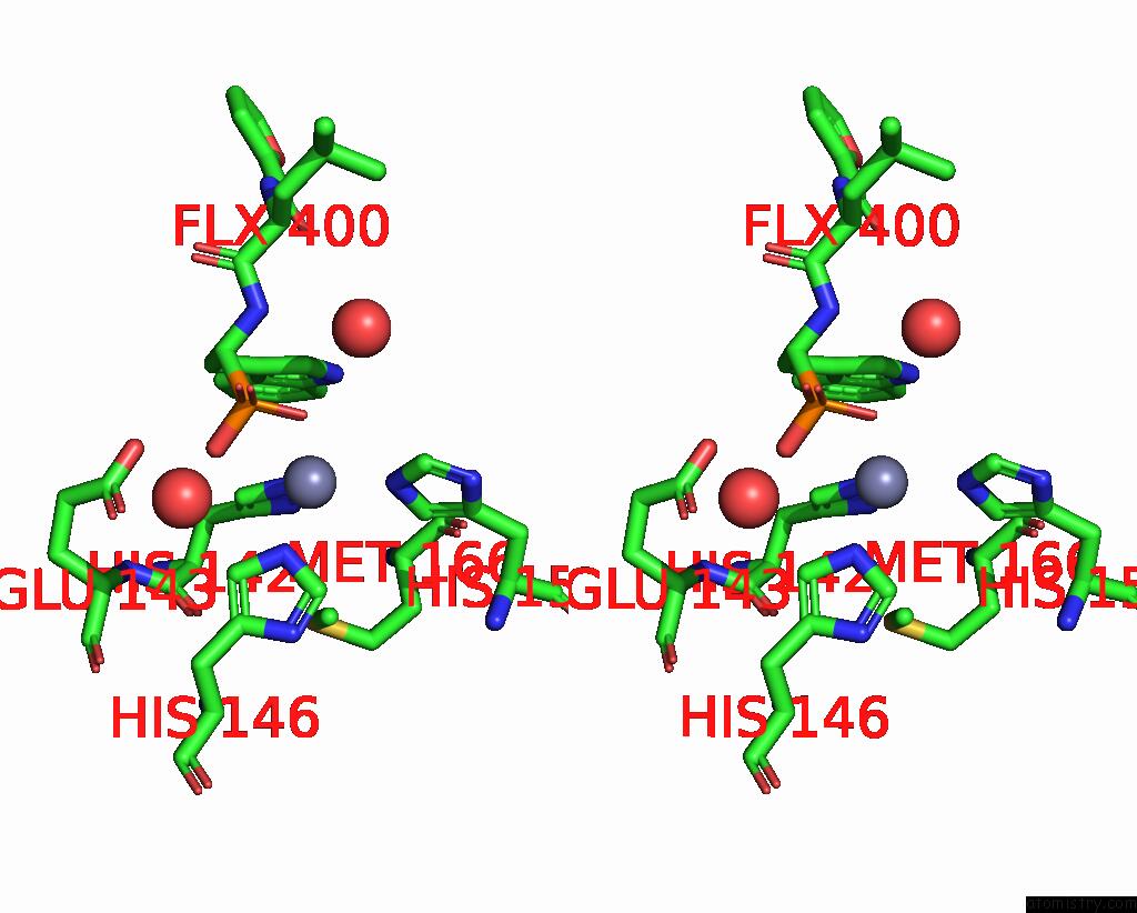
Stereo pair view
Mono view
Stereo pair view
A full contact list of Zinc with other atoms in the Zn binding
site number 1 of Adamalysin II with Phosphonate Inhibitor within 5.0Å range:
|
Reference:
M.Cirilli,
C.Gallina,
E.Gavuzzo,
C.Giordano,
F.X.Gomis-Ruth,
B.Gorini,
L.F.Kress,
F.Mazza,
M.P.Paradisi,
G.Pochetti,
V.Politi.
2 Angstrom X-Ray Structure of Adamalysin II Complexed with A Peptide Phosphonate Inhibitor Adopting A Retro-Binding Mode. Febs Lett. V. 418 319 1997.
ISSN: ISSN 0014-5793
PubMed: 9428736
DOI: 10.1016/S0014-5793(97)01401-4
Page generated: Sat Oct 26 19:14:39 2024
ISSN: ISSN 0014-5793
PubMed: 9428736
DOI: 10.1016/S0014-5793(97)01401-4
Last articles
Zn in 9MJ5Zn in 9HNW
Zn in 9G0L
Zn in 9FNE
Zn in 9DZN
Zn in 9E0I
Zn in 9D32
Zn in 9DAK
Zn in 8ZXC
Zn in 8ZUF