Zinc »
PDB 4a3i-4ac1 »
4a7g »
Zinc in PDB 4a7g: Structure of Human I113T SOD1 Mutant Complexed with 4-Methylpiperazin- 1-Yl)Quinazoline in the P21 Space Group.
Enzymatic activity of Structure of Human I113T SOD1 Mutant Complexed with 4-Methylpiperazin- 1-Yl)Quinazoline in the P21 Space Group.
All present enzymatic activity of Structure of Human I113T SOD1 Mutant Complexed with 4-Methylpiperazin- 1-Yl)Quinazoline in the P21 Space Group.:
1.15.1.1;
1.15.1.1;
Protein crystallography data
The structure of Structure of Human I113T SOD1 Mutant Complexed with 4-Methylpiperazin- 1-Yl)Quinazoline in the P21 Space Group., PDB code: 4a7g
was solved by
G.S.A.Wright,
N.M.Kershaw,
R.Sharma,
S.V.Antonyuk,
R.W.Strange,
N.G.Berry,
P.M.O'neil,
S.S.Hasnain,
with X-Ray Crystallography technique. A brief refinement statistics is given in the table below:
| Resolution Low / High (Å) | 48.33 / 1.24 |
| Space group | P 1 21 1 |
| Cell size a, b, c (Å), α, β, γ (°) | 38.793, 68.352, 49.920, 90.00, 104.49, 90.00 |
| R / Rfree (%) | 13.9 / 17.5 |
Other elements in 4a7g:
The structure of Structure of Human I113T SOD1 Mutant Complexed with 4-Methylpiperazin- 1-Yl)Quinazoline in the P21 Space Group. also contains other interesting chemical elements:
| Copper | (Cu) | 3 atoms |
Zinc Binding Sites:
The binding sites of Zinc atom in the Structure of Human I113T SOD1 Mutant Complexed with 4-Methylpiperazin- 1-Yl)Quinazoline in the P21 Space Group.
(pdb code 4a7g). This binding sites where shown within
5.0 Angstroms radius around Zinc atom.
In total 2 binding sites of Zinc where determined in the Structure of Human I113T SOD1 Mutant Complexed with 4-Methylpiperazin- 1-Yl)Quinazoline in the P21 Space Group., PDB code: 4a7g:
Jump to Zinc binding site number: 1; 2;
In total 2 binding sites of Zinc where determined in the Structure of Human I113T SOD1 Mutant Complexed with 4-Methylpiperazin- 1-Yl)Quinazoline in the P21 Space Group., PDB code: 4a7g:
Jump to Zinc binding site number: 1; 2;
Zinc binding site 1 out of 2 in 4a7g
Go back to
Zinc binding site 1 out
of 2 in the Structure of Human I113T SOD1 Mutant Complexed with 4-Methylpiperazin- 1-Yl)Quinazoline in the P21 Space Group.
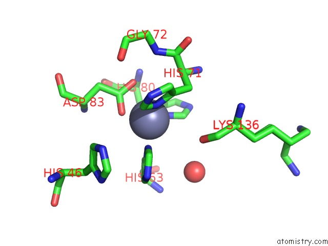
Mono view
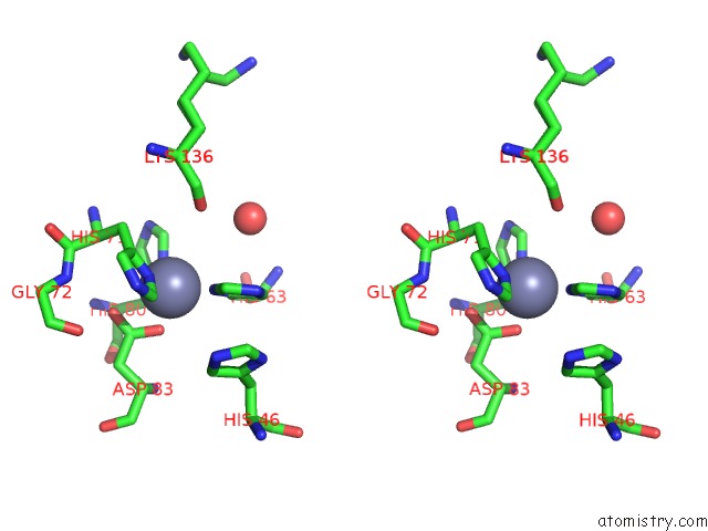
Stereo pair view
Mono view
Stereo pair view
A full contact list of Zinc with other atoms in the Zn binding
site number 1 of Structure of Human I113T SOD1 Mutant Complexed with 4-Methylpiperazin- 1-Yl)Quinazoline in the P21 Space Group. within 5.0Å range:
|
Zinc binding site 2 out of 2 in 4a7g
Go back to
Zinc binding site 2 out
of 2 in the Structure of Human I113T SOD1 Mutant Complexed with 4-Methylpiperazin- 1-Yl)Quinazoline in the P21 Space Group.
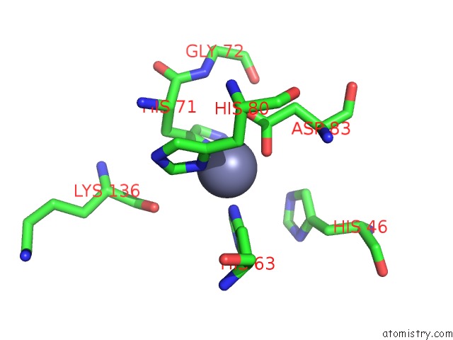
Mono view
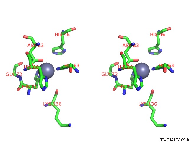
Stereo pair view
Mono view
Stereo pair view
A full contact list of Zinc with other atoms in the Zn binding
site number 2 of Structure of Human I113T SOD1 Mutant Complexed with 4-Methylpiperazin- 1-Yl)Quinazoline in the P21 Space Group. within 5.0Å range:
|
Reference:
N.M.Kershaw,
G.S.Wright,
R.Sharma,
S.V.Antonyuk,
R.W.Strange,
N.G.Berry,
P.M.O'neill,
S.S.Hasnain.
X-Ray Crystallography and Computational Docking For the Detection and Development of Protein-Ligand Interactions. Curr.Med.Chem. V. 20 569 2013.
ISSN: ISSN 0929-8673
PubMed: 23278398
DOI: 10.2174/0929867311320040008
Page generated: Sat Oct 26 19:04:39 2024
ISSN: ISSN 0929-8673
PubMed: 23278398
DOI: 10.2174/0929867311320040008
Last articles
Zn in 9MJ5Zn in 9HNW
Zn in 9G0L
Zn in 9FNE
Zn in 9DZN
Zn in 9E0I
Zn in 9D32
Zn in 9DAK
Zn in 8ZXC
Zn in 8ZUF